
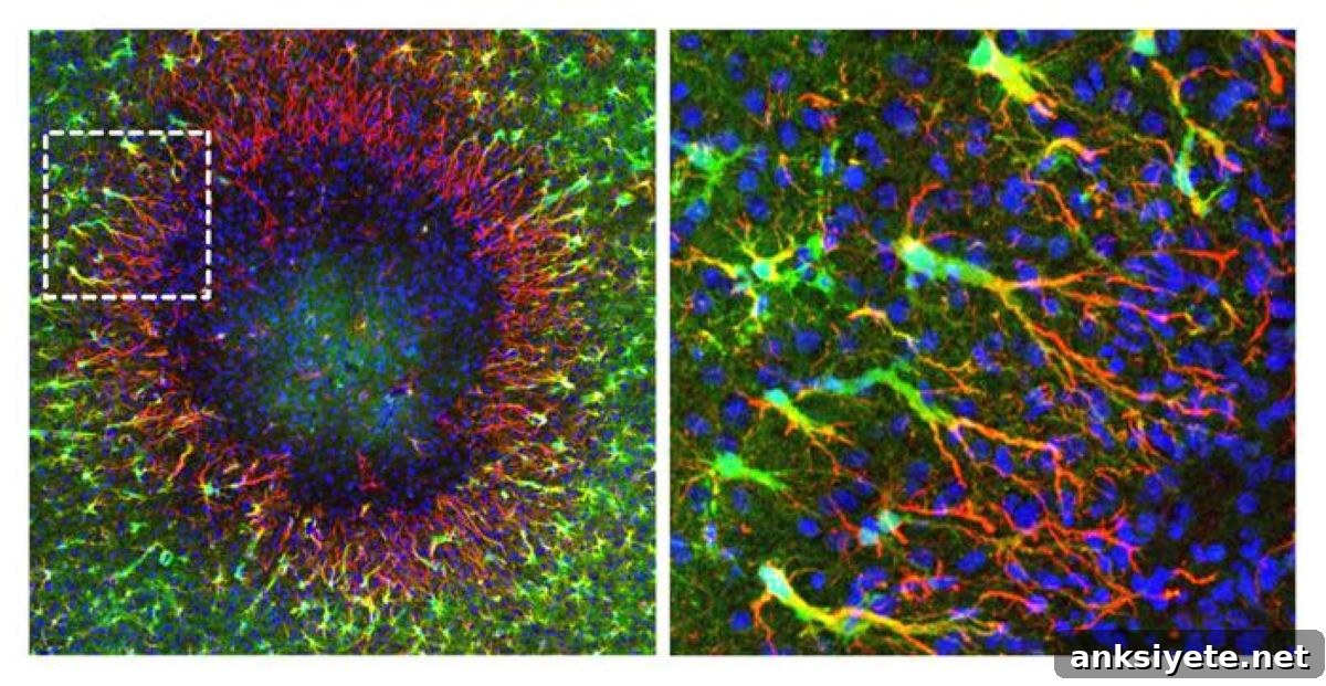
Summary: Researchers have uncovered a previously unknown mechanism by which the adult mammalian brain can repair localized tissue damage. Using longitudinal in vivo two-photon imaging and spatial gene expression mapping in mouse models, the team at the University of Zurich identified a distinct population of “regenerative” astrocytes that repopulate lesions by transporting newly formed cell nuclei through long astrocytic processes. This discovery—highlighting nuclear migration as a driver of astrocyte network reconstruction—points to molecular pathways that could be targeted to enhance recovery after traumatic brain injury and autoimmune astrocytopathies such as neuromyelitis optica spectrum disorder (NMOSD).
Astrocytes are star-shaped glial cells that support neuronal function by supplying nutrients, maintaining extracellular ion balance, and regulating local blood flow. When astrocytes are lost after injury or in autoimmune diseases, the resulting disruption of local tissue homeostasis has been thought to be only partially reversible in the adult brain. This study overturns that assumption by demonstrating an active regenerative program in perilesional astrocytes that restores the astrocyte network and tissue architecture.
Key Facts
- New paradigm for glial regeneration: The adult central nervous system can replace lost astrocytes and rebuild local tissue structure through a specialized repair process, expanding current understanding of astroglial plasticity.
- Nuclear migration mechanism: Regenerative astrocytes positioned at the lesion boundary divide and send the nuclei of daughter cells along extended cellular processes into the depleted lesion core, rather than relying solely on cell body migration or local division within the lesion.
- Restoration of essential functions: By reconstructing astrocyte networks, this process helps re-establish critical homeostatic roles—nutrient delivery, blood flow regulation via astrocytic end-feet, and extracellular ionic control—supporting neuronal survival and function.
- Clinical relevance: The mechanism has potential therapeutic implications for traumatic brain injury, stroke, and autoimmune conditions such as NMOSD, where autoantibodies selectively attack astrocytes.
- Drug discovery targets: The researchers identified transiently activated genes and signaling pathways that accompany nuclear migration and lesion repopulation, offering candidate targets for drugs that could accelerate or enhance brain repair.
Source: University of Zurich
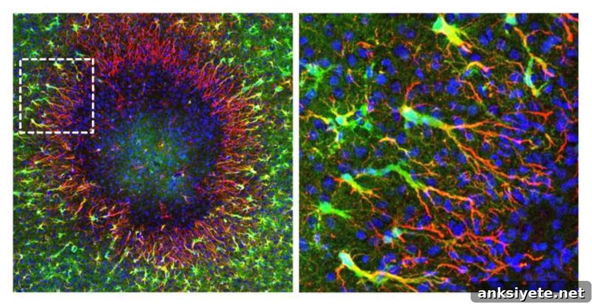
Regenerative astrocytes repair damaged tissue
A research team led by Bruno Weber at the Institute of Pharmacology and Toxicology, University of Zurich, discovered a distinct subset of astrocytes that mediate lesion repopulation in living mice. Co-lead authors Marina Herwerth and Matthias Wyss used repeated two-photon imaging to follow the same brain regions over weeks and combined these observations with spatial transcriptional profiling to define the molecular programs activated during repair.
Instead of the classical model in which cells either divide locally or migrate as whole cell bodies into a lesion, the regenerative astrocytes remain anchored at the lesion perimeter. They proliferate and generate daughter-cell nuclei that move along elongated astrocytic processes into the empty territory, gradually reoccupying the lesion and restoring a functional astrocyte network. This nuclear translocation is accompanied by pronounced structural remodeling—process polarization, transient multinucleated astrocyte states, and progressive displacement of nuclei into previously unoccupied domains.
Therapeutic starting points
Mapping gene expression over time revealed an injury-associated molecular response that resolves as the astrocyte network is re-established. The researchers cataloged numerous genes and signaling pathways transiently upregulated during nuclear migration and tissue repopulation. These pathways provide logical starting points for experimental interventions aimed at enhancing endogenous regeneration after astrocyte loss, with potential application in neurotrauma and astrocytopathies including NMOSD.
According to the authors, selectively activating or modulating these regenerative mechanisms could improve structural and functional recovery after lesions that deplete astrocytes. The findings broaden our understanding of adult brain plasticity and highlight previously unrecognized cellular behaviors that contribute to tissue restoration.
Key Questions Answered:
A: Regenerative astrocytes stay at the lesion boundary, divide there, and transport the nuclei of daughter cells through long processes directly into the depleted area. This nuclear migration repopulates the lesion without whole-cell body migration into the core.
A: Astrocyte loss can result from traumatic brain injury, stroke, and neuroinflammatory or autoimmune disorders—most notably Neuromyelitis Optica Spectrum Disorder (NMOSD), where autoantibodies target and destroy astrocytes.
A: The team used in vivo two-photon microscopy to image living mouse cortex repeatedly over several weeks, enabling direct visualization of astrocytic process dynamics, nuclear movement, and concurrent spatial changes in gene expression during lesion repopulation.
Editorial Notes:
- Article edited by a Neuroscience News editor.
- Full journal paper reviewed.
- Additional explanatory context added by editorial staff.
About this neuroscience research news
Author: Kurt Bodenmueller
Source: University of Zurich
Contact: Kurt Bodenmueller – University of Zurich
Image credit: Institute of Pharmacology and Toxicology, University of Zurich
Original Research: Open access. “Focal astrocyte loss reveals nuclear translocation during lesion repopulation” by Marina Herwerth, Matthias T. Wyss, Nicola B. Schmid, Anna Lasne, Jacqueline Condrau, Luca Ravotto, José María Mateos Melero, Andres Kaech, Gustav Bredell, Carolina Thomas, Rachel Kim, Petra Kukanja, Vladyslav L. Korobeynyk, Christine Stadelmann, Thomas Misgeld, Jeffrey L. Bennett, Sebastian Jessberger, Aiman S. Saab, Shane A. Liddelow & Bruno Weber. Nature Neuroscience. DOI: 10.1038/s41593-026-02354-5
Abstract
Focal astrocyte loss reveals nuclear translocation during lesion repopulation
Astrocyte loss in localized brain lesions disrupts tissue homeostasis and can impair neuronal support. While astrocytes bordering many lesions enter reactive states that do not fully restore the astrocyte network, the response to spatially confined astrocyte depletion has been unclear. Using longitudinal in vivo two-photon microscopy combined with spatiotemporal transcriptional profiling, this study examines astrocyte behavior after focal aquaporin-4 antibody–mediated ablation in adult mouse somatosensory cortex—a model relevant to neuromyelitis optica spectrum disorder.
Perilesional astrocytes undergo extensive structural remodeling during repopulation: they proliferate, often exhibit transient multinucleated states, extend polarized processes into the depleted area, and progressively displace nuclei into previously unoccupied territories. Spatial transcriptomics identify an injury-associated molecular program that resolves as the astrocyte network is reestablished. Together, these results define the spatiotemporal dynamics of astrocyte regeneration after focal loss and extend current understanding of astroglial plasticity in the adult brain.