
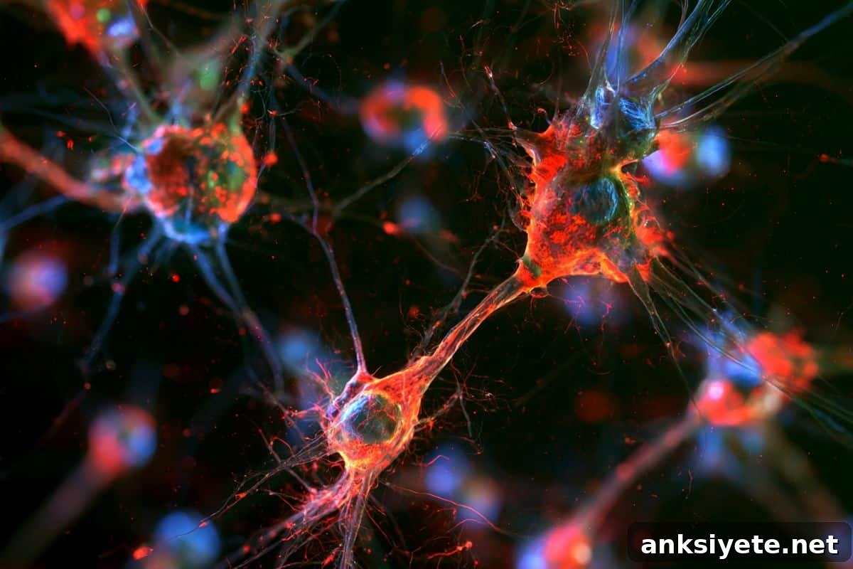
Summary: Researchers have discovered that aging drives a substantial and previously unrecognized migration of bone marrow–derived immune cells from the blood into the human brain. This finding overturns the long-standing view that the brain’s immune compartment is entirely maintained by resident microglia established before birth and isolated from peripheral influence.
Key Facts
- Shifts long-held neuroimmunology assumptions: The study challenges the idea that microglia in the brain self-renew in isolation, showing instead that aging human brains receive ongoing immune cell input from bone marrow-derived blood stem cells.
- Lineage tracing via somatic DNA mutations: Scientists compared somatic mutations detected in blood stem cells and brain tissue to provide genetic evidence that many microglia-like cells in aged human brains share progenitors with circulating blood immune cells.
- Human-specific aging feature: This migration and subsequent conversion of peripheral immune cells into microglia-like cells appears to be unique to humans and was not observed in standard laboratory models such as mice or non-human primates.
- Connection to clonal hematopoiesis and Alzheimer’s risk: The research builds on prior observations linking certain mutated blood stem cell clones (clonal hematopoiesis) to altered Alzheimer’s risk, indicating that peripheral immune cells actively infiltrate the brain and can change disease trajectories.
- Therapeutic potential for brain immunotherapies: Because engineered peripheral immune cells naturally home to the aging human brain, the finding opens the possibility of designing cell-based therapies to clear amyloid-beta or tau aggregates before symptoms appear.
Source: Stanford University
Background: The brain’s immune environment has traditionally been regarded as largely separate from the rest of the body, maintained by microglia seeded during embryonic development and protected by the blood-brain barrier. New evidence from Stanford researchers now shows significant infiltration of peripheral immune cells into the brain with aging, a discovery that reshapes our understanding of brain immunity and suggests new therapeutic strategies.
The study, supported in part by the Knight Initiative for Brain Resilience at the Wu Tsai Neurosciences Institute, was published July 30, 2026 in the journal Nature.
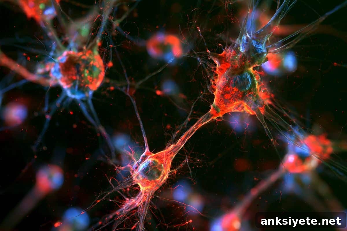
“We usually think of the brain as a closed system,” said Julia Belk, a postdoctoral scholar in pathology at Stanford Medicine and first author on the study. “What we found is that a substantial number of immune cells enter the human brain during aging.”
How the discovery unfolded
Julia Belk’s interdisciplinary background in computer science and biomedical training equipped her to combine genetic analysis with cellular biology. Collaborating with senior author Siddhartha Jaiswal, an associate professor of pathology, the team examined genetic data across thousands of people—some followed for decades—and previously observed that specific blood stem cell clones bearing somatic mutations correlated with reduced Alzheimer’s risk. That pattern suggested a possible interaction between circulating immune cells and the brain.
While the clonal hematopoiesis mutations responsible for those protective clones affect only a subset of individuals, the team asked whether peripheral immune cell infiltration into the brain might be a broader feature of human aging. To test this, they needed direct evidence that immune cells inside the brain arose from blood stem cells rather than from embryonically seeded microglia.
Tracing a cell’s lineage using DNA
The researchers used donated samples of blood and post-mortem brain tissue from the Stanford Rapid Autopsy Center and the University of Washington Alzheimer’s Disease Sequencing Project. Their method relied on the natural accumulation of harmless somatic mutations in blood stem cells over time. Because the progeny of a stem cell inherit those mutations, identical mutation patterns in cells from two compartments strongly indicate a shared origin.
By sequencing DNA from immune cells in both blood and brain tissue, the team identified matching somatic mutations, demonstrating that marrow-derived cells had migrated into the brain as early as middle age. Single-cell analyses, including mitochondrial DNA lineage tracing, showed that these infiltrating cells adopt microglia-like identities and in some individuals form a substantial proportion of the microglial pool. Comparative work indicates this pattern is not replicated in common animal models, highlighting its human specificity.
Implications for neurodegeneration and therapies
The discovery carries several potential implications for aging and neurodegenerative disease research. First, it suggests that the life history of an individual’s blood stem cells—including the expansion of specific mutant clones—can shape the brain’s immune landscape and influence disease risk.
Second, because peripheral immune cells can naturally access the aging brain and become microglia-like, they represent a deliverable platform for engineered immunotherapies. Researchers could potentially modify a patient’s circulating immune cells to target and clear toxic protein aggregates such as amyloid-beta and tau before neurodegeneration advances.
“Now that we know these cells can enter the brain, we can explore engineering strategies that harness peripheral immune cells for protective brain functions,” Belk said. She also emphasized that this appears to be a uniquely human aging feature, which explains why the phenomenon was not apparent in standard animal experiments.
Key Questions Answered
A: The team sequenced DNA from peripheral blood and brain tissue to identify matching somatic mutations. These shared genetic signatures provide lineage evidence that some brain immune cells are descendants of mutated blood stem cells.
A: Detailed comparisons indicate this infiltration and conversion into microglia-like cells is a feature observed in aging humans but not in commonly used model organisms such as mice or non-human primates.
A: Because peripheral immune cells can access the aging brain, they could be engineered to perform targeted protective tasks—such as degrading amyloid-beta or tau aggregates—offering a potential preventive approach to neurodegeneration.
Editorial Notes:
- This article was edited by a Neuroscience News editor.
- The referenced journal paper was reviewed in full.
- Additional context was added by editorial staff.
About this aging and neuroscience research news
Author: Nathan Collins
Source: Stanford University
Contact: Nathan Collins – Stanford University
Image credit: Neuroscience News
Original Research: Open access. “Somatic mutations reveal the ontogeny of microglia in human aging” by Julia A. Belk et al., published in Nature. DOI: 10.1038/s41586-026-10939-0
Abstract
Somatic mutations reveal the ontogeny of microglia in human aging
Microglia are the resident macrophages of the central nervous system. In mice, microglia are seeded during embryogenesis and maintained with little contribution from adult hematopoiesis. The origins of human microglia have been less clear, but recent evidence suggested marrow-derived cells might contribute in some people.
To study human microglial ontogeny, the authors developed an approach that uses accumulated somatic mutations as natural clonal labels to trace marrow-derived cell infiltration into the human brain. Applying this method to samples from aged individuals, they found evidence of marrow-derived cell influx in all examined cases. Single-cell analysis, including mitochondrial DNA lineage tracing, showed that these infiltrating cells resemble microglia and can represent a substantial fraction of the microglial pool.
Analysis of cohort data also revealed a protective association between many forms of clonal hematopoiesis and Alzheimer’s disease. Together, the results uncover a widespread influx of myeloid cells into the healthy aging human brain that contributes to the microglial population and becomes common with age.