
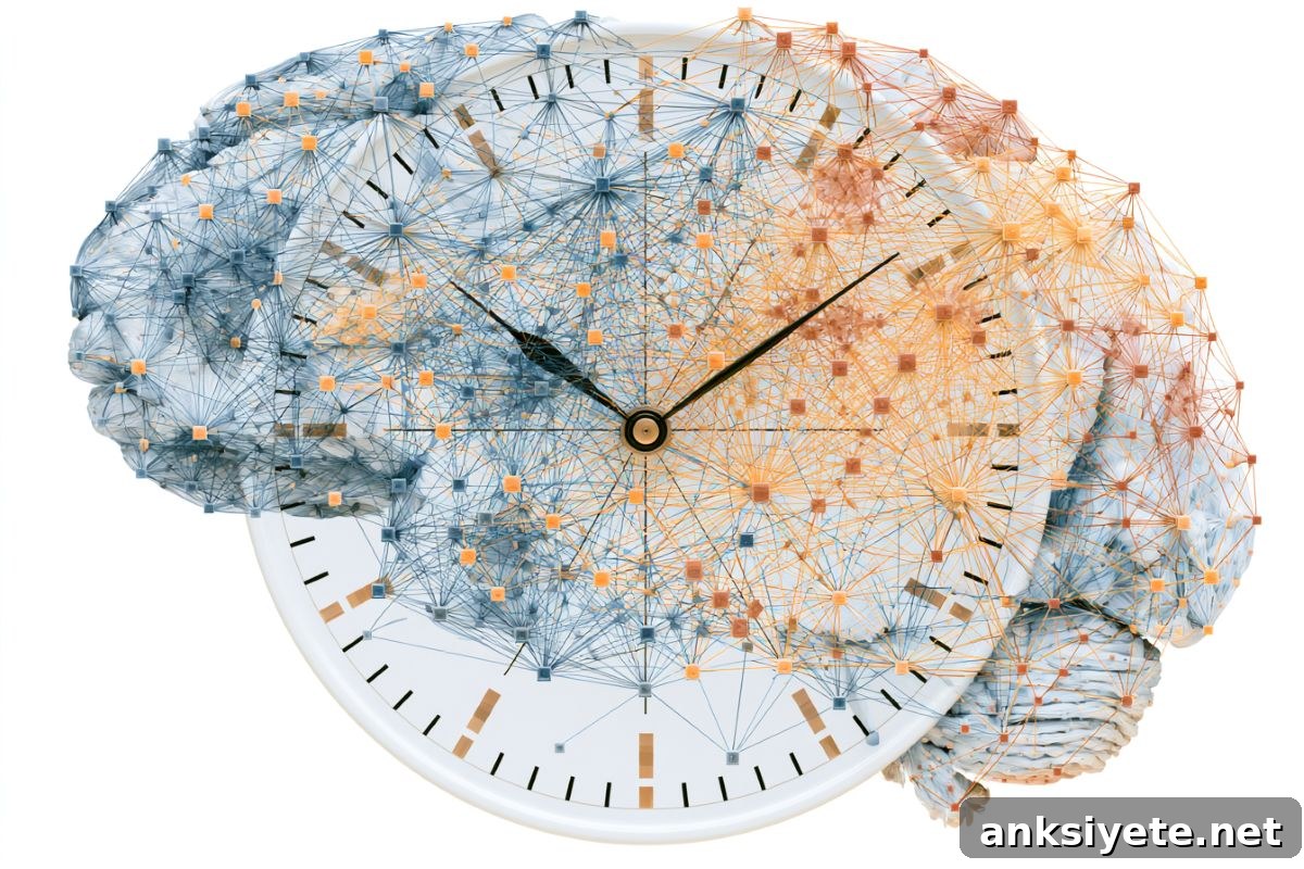
Summary: Using wide-field two-photon calcium imaging while mice performed a temporal expectation task, researchers simultaneously recorded thousands of neurons in the secondary motor cortex (M2) and the posterior parietal cortex (PPC).
The team found that M2 and PPC are neither perfectly synchronized nor fully independent. Instead, they operate in a balanced regime where sparse inter-regional connections provide enough coupling to keep timing broadly aligned, while widespread neural fluctuations preserve local flexibility and allow each region to represent time independently when needed.
Key Facts
- Sequential population dynamics: Both M2 and PPC represent elapsed time through sequential patterns of neuronal activation, with distinct ensembles firing in succession to mark different moments.
- Two distinct error modes in timing: Decoding analyses revealed two types of timing errors during interval prediction:
- Correlated drift: Trials in which both M2 and PPC misestimated elapsed time in the same direction, indicating a shared deviation.
- Independent drift: Trials in which one region drifted off while the other remained accurate, indicating local autonomy.
- Sparse coupling mechanism: A twin recurrent neural network (RNN) model showed that sparse inter-regional connectivity gently tethers the two areas, supporting overall coherence without forcing identical timing states.
- Global fluctuations preserve autonomy: Shared, high-variance neural fluctuations act as a counterweight to excessive synchronization, enabling each cortical area to flexibly track separate information streams when the task demands it.
- Broader implications: This framework clarifies how distributed cortical systems can balance coordinated behavior with local flexibility, with potential relevance for understanding neuropsychiatric disorders and for designing brain-inspired AI and multi-agent control systems.
Source: Institute of Science Tokyo
Timekeeping is a continuous, largely implicit function of the brain. The ability to sense elapsed time underlies movement, planning, working memory, decision-making and learning. Many higher-order cortical areas, particularly frontal and parietal regions, encode temporal information. When multiple regions represent time simultaneously, a key question is how they remain coordinated without losing the flexibility to act independently.
Researchers led by Associate Professor Riichiro Hira in the Department of Physiology and Cell Biology at the Institute of Science Tokyo approached this question by measuring large-scale neural activity across two frontal-parietal areas while mice performed a carefully designed timing task. Their study, published in Volume 17 of Nature Communications on June 11, 2026, focused on the secondary motor cortex and the posterior parietal cortex during an alternating-interval reward task.
Mice were trained to anticipate rewards that alternated between 6-second and 12-second intervals. After learning the pattern, animals reliably predicted both short and long intervals. During task performance the team applied mesoscale, wide-field two-photon calcium imaging to record activity from thousands of neurons in M2 and PPC simultaneously, enabling trial-by-trial comparisons of temporal representations across regions.
Both regions displayed high-dimensional sequential activity: different neurons peaked at successive moments across each interval, forming population trajectories that encoded elapsed time. By decoding these population patterns, the researchers could estimate the moment in time represented by each region on each trial. This decoding revealed two complementary error modes — correlated drifts shared by both regions and independent drifts confined to one region — demonstrating that the fronto-parietal network can operate coherently or independently depending on internal and external factors.
To identify circuit mechanisms that produce these mixed modes, the team developed a computational model composed of twin RNNs with sparse inter-network connections and shared, high-variance noise. This model reproduced the experimental observations: sparse coupling promoted broad synchronization, while shared global fluctuations and intrinsic dynamics allowed each network to retain local timing variability. Analyses of communication subspaces and perturbations further showed that different shared dimensions selectively support either coherent or independent temporal computation.
In short, the brain appears to use a hybrid strategy: weak, specific inter-area coupling keeps regions loosely aligned, and widespread fluctuations prevent over-synchronization so each area can maintain flexible, local computations. This balance enables robust, distributed temporal processing across the frontal-parietal network.
“Our results suggest a circuit-level design principle for balancing stability and flexibility in distributed timing,” says Riichiro Hira. “This insight may inform models of cognitive dysfunction where inter-regional coordination breaks down and could inspire AI and robotics architectures that require both unified goals and modular flexibility.”
Key Questions Answered:
A: The brain appears to use a hybrid approach. Rather than depending on a single master clock or fully independent timers, loosely connected local clocks across cortical regions maintain their own sequential timing patterns while sharing just enough coupling to stay aligned when required.
A: Wide-field two-photon calcium imaging enables simultaneous visualization of activity from thousands of individual neurons across large cortical areas with cellular resolution. It was essential here because the study required concurrent recordings from M2 and PPC to detect trial-by-trial timing errors and to compare population dynamics across regions in real time.
A: Many AI and robotic systems must balance unified control with module-level flexibility. The sparse coupling model offers a design principle: maintain weak, targeted connections between modules to preserve global coordination while allowing local modules to process information independently, improving resilience and adaptability.
Editorial Notes:
- Article edited by a Neuroscience News editor.
- Original journal paper reviewed in full by editorial staff.
- Additional explanatory context added by the newsroom.
About this neuroscience research news
Author: Miki Yamaoka
Source: Institute of Science Tokyo
Contact: Miki Yamaoka – Institute of Science Tokyo
Image: Image credit: Neuroscience News
Original Research: Open access.
“Independence and coherence in temporal sequence computation across the fronto-parietal network” by Hiroto Imamura, Fumiya Imamura, Reiko Hira, Yoshikazu Isomura & Riichiro Hira. Nature Communications
DOI: 10.1038/s41467-026-73999-w
Abstract
Independence and coherence in temporal sequence computation across the fronto-parietal network
Temporal processing depends on distributed and coordinated cortical dynamics, but how multiple brain areas flexibly switch between coherent and independent temporal representations has been unclear. Using mesoscale two-photon calcium imaging, the authors simultaneously recorded neuronal populations in the secondary motor cortex and posterior parietal cortex of mice performing an alternating-interval timing task. Both areas encoded elapsed time through similar high-dimensional sequential activity, and decoding analyses revealed both coherent temporal errors shared across areas and independent errors confined to a single area.
Communication-subspace analyses showed that temporal information spreads across multiple low-variance shared dimensions, while the dominant shared dimension preferentially encoded behavior. A twin recurrent neural network model with sparse inter-network coupling and shared high-variance noise reproduced these experimental findings. Perturbation and Lyapunov exponent analyses indicated that different shared subspaces selectively promote coherent or independent modes. These results demonstrate how sparse coupling combined with shared global fluctuations supports robust yet flexible temporal computation across the fronto-parietal network.