
Summary: A new study shows that branched O-mannose glycans produced by the brain-specific enzyme MGAT5B are essential to maintain the narrow architecture of the nodes of Ranvier. Mice lacking MGAT5B develop abnormally widened axonal nodes in white matter, which disrupts saltatory conduction, slows nerve propagation, and impairs motor coordination.
The research team traced the defect to altered glycosylation of neurofascin 186 (NF186), which weakens its interaction with Contactin 1, establishing glycan branching as an active regulator of myelinated nerve fiber stability.
Key Facts
- Localized Nodal Structural Defect: Deletion of the MGAT5B enzyme in mice produced a precise anatomical change: the nodes of Ranvier in brain white matter were significantly widened.
- Disrupted Saltatory Conduction: Nodes of Ranvier are microscopic relay gaps in the myelin sheath that enable action potentials to jump rapidly down the axon. When these gaps widen, electrical impulses leak and conduction becomes slower and more variable.
- Motor Performance Declines: The electrophysiological slowdown correlated with measurable motor deficits: MGAT5B knockout mice performed worse on balance and coordination tests than controls.
- Molecular Target — NF186: The team identified neurofascin 186 (NF186) as a critical node-organizing glycoprotein. MGAT5B attaches branched O-mannose glycans to NF186, and these glycans regulate NF186’s interaction with Contactin 1 to preserve a narrow nodal gap required for efficient conduction.
- Neuron-Specific Action Confirmed: Restoring MGAT5B expression specifically in neurons rescued the nodal abnormalities, demonstrating that the enzyme functions cell-autonomously within axons to maintain node structure.
- Relevance to Demyelinating Conditions: By revealing how glycan branching supports node integrity, the study points to a new mechanism to explore in demyelinating and white matter diseases, such as multiple sclerosis, where nodal structure and conduction are compromised.
Source: IGCORE
Sugarcoating isn’t only about flavor — in the brain, complex sugar chains decorate key proteins and determine how they function.
Researchers at Gifu University’s Institute for Glyco-core Research (iGCORE) discovered that a specific class of brain glycans — branched O-mannose glycans generated by MGAT5B — is required to maintain the tiny nodal structures that allow rapid and reliable electrical signaling.
The study was published in Communications Biology on July 13.
Glycans are sugar chains attached to proteins that influence protein folding, stability, and interactions. Although O-mannose glycans are known to be important in muscle, their functions in the brain have been less clear despite their abundance there. Prior links between O-mannose defects and neurological disorders motivated the current investigation.
To examine MGAT5B’s role, the team generated Mgat5b knockout mice and combined biochemical analyses with electrophysiology and behavioral testing. They observed that loss of MGAT5B produced marked broadening of nodes of Ranvier in brain white matter. Electrophysiological measurements showed delayed and more variable axonal conduction in knockout mice, and behavioral tests revealed impaired motor coordination.
Biochemical and glycoproteomic experiments implicated NF186: MGAT5B modifies NF186 with branched O-mannose glycans, and these modifications negatively regulate the interaction between NF186 and Contactin 1. Proper regulation of this molecular interaction preserves the narrow nodal architecture required for fast, reliable saltatory conduction.
Importantly, reintroducing MGAT5B specifically in neurons rescued the nodal defects in knockout animals, supporting a direct, cell-autonomous role for the enzyme in node organization.
These results identify branched O-mannose glycans as critical regulators of node of Ranvier formation and function, providing a physiological explanation for the presence of these brain-specific sugar structures and opening new avenues to study how glycosylation defects might contribute to myelin-related disorders.
The authors note that questions remain: the exact molecular mechanism by which O-mannose glycan branching determines node width is still unresolved, and the detailed structures of the glycans attached to NF186 require further characterization. Future work will explore whether defects in these sugar modifications contribute to diseases that affect myelin or nerve conduction.
Funding
- FOREST program nos. JPMJFR2145 and JPMJFR215Z from the Japan Science and Technology Agency (JST)
- Grant-in-Aid for Scientific Research (B) no. 24K02222; Grants-in-Aid for Scientific Research (C) no. JP23K06302; Core-to-Core Program no. JPJSCCA202000007; J-PEAKS program no. JPJS00420230009 from the Japan Society for the Promotion of Science (JSPS)
- AMED-CREST grant no. JP23gm1410011 from the Japan Agency for Medical Research and Development (AMED)
- Human Glycome Atlas Project (HGA) from the Japanese Ministry of Education, Culture, Sports, Science and Technology (MEXT)
Key Questions Answered:
A: Proteins are commonly modified by complex, branching chains of sugar molecules called glycans in a process known as glycosylation. These sugar chains influence protein folding, stability, and interactions with other molecules. In the nervous system, glycans help proteins assemble and anchor into the precise structures—like the nodes of Ranvier—needed for stable, high-speed nerve conduction.
A: Axons are insulated by myelin, which prevents continuous conduction along the membrane. Tiny gaps in that insulation—the nodes of Ranvier—concentrate voltage-gated ion channels and allow action potentials to jump from node to node in a process called saltatory conduction. This arrangement enables rapid, energy-efficient signal propagation; if the nodes widen or their molecular architecture is disrupted, conduction slows and becomes less reliable.
A: The finding that MGAT5B-dependent glycan branching actively maintains node structure suggests new therapeutic strategies for white matter and demyelinating conditions. Rather than focusing solely on restoring myelin, targeting glycosylation pathways that preserve nodal organization could provide alternative ways to protect or restore nerve conduction in disorders such as multiple sclerosis.
Editorial Notes:
- This article was edited by a Neuroscience News editor.
- The journal paper was reviewed in full.
- Additional context added by editorial staff.
About this neuroscience research news
Author: Shinji Ito
Source: iGCORE
Contact: Shinji Ito – iGCORE
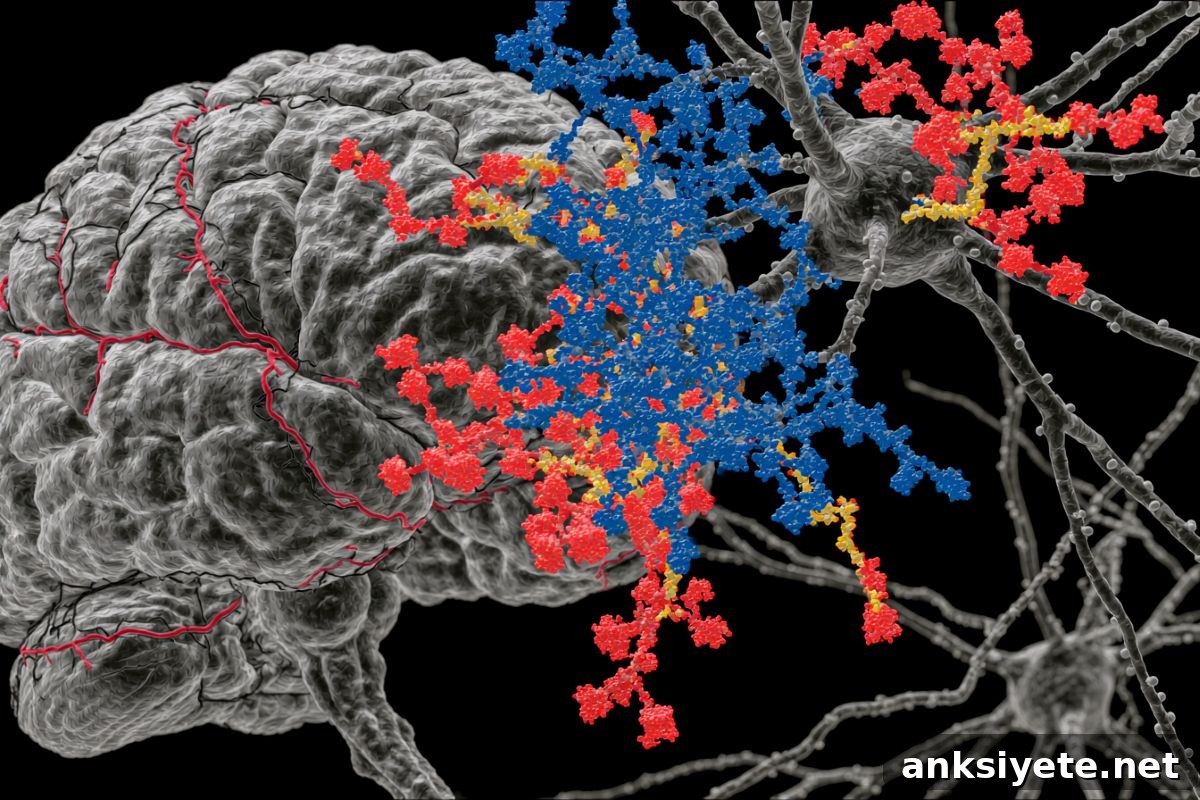
Image: The image is credited to Neuroscience News
Original Research: Open access. “Branching of O-mannose glycans regulates node of Ranvier organization and saltatory conduction” by Shu Tomita, Taichi Nakaishi, Toshiyuki Ishii, Kazuya Ono, Honoka Fujimori, Misuzu Hashimoto, Shiho Ohno, Yoshiki Yamaguchi, Masamitsu Shimazawa, Miyako Nakano, Daisuke Kato & Yasuhiko Kizuka. Communications Biology. DOI: 10.1038/s42003-026-10622-0
Abstract
Branching of O-mannose glycans regulates node of Ranvier organization and saltatory conduction
The myelin sheath of axons is organized into domain structures with nodes of Ranvier that facilitate saltatory conduction. A brain-specific glycosyltransferase, MGAT5B, catalyzes β1,6-GlcNAc branching of O-mannose glycans and is required for node integrity. Mgat5b knockout mice displayed broadening of nodes in brain white matter.
Electrophysiological analysis showed delayed and variable axonal conduction in Mgat5b knockout mice, indicating the importance of branched O-mannose glycans in node morphology and function. Biochemical and glycoproteomic analyses demonstrated that MGAT5B modifies the glycans of the node-organizing glycoprotein NF186, and that branched O-mannose glycans negatively regulate the interaction between NF186 and Contactin 1.
Neuron-specific restoration of MGAT5B in knockout mice rescued the nodal defects, indicating a cell-autonomous role for MGAT5B in node organization. These findings highlight a glycan-mediated mechanism for maintaining node structure and function.