
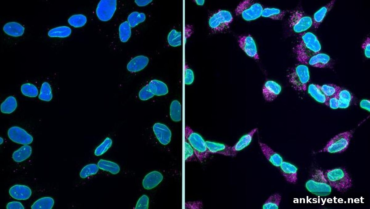
Summary: For decades, researchers have linked toxic protein build-up to neurodegenerative diseases such as Alzheimer’s disease, frontotemporal dementia (FTD), and amyotrophic lateral sclerosis (ALS). Yet how these aggregates drive widespread neuronal loss remained unclear. Conventional forms of programmed cell death like apoptosis do not fully explain the large-scale disappearance of neurons observed in dementia. New research now identifies a distinct cell-death process, called karyoptosis, that connects proteotoxic stress to nuclear collapse and neuron loss.
Karyoptosis is a chemically driven sequence of events initiated by accumulated misfolded proteins inside neurons. This cascade destabilizes the nuclear envelope, causing the nucleus to shrink, deform, and eventually disintegrate. By analyzing more than 3,000 cells from post-mortem human brains, researchers report that this mechanism actively contributes to neuronal death in common dementias and offers a focused target for therapies aimed at preserving brain structure and function.
Key Facts
- Karyoptosis signature: Defined by a specific chain of biochemical reactions in which toxic intracellular protein aggregates compromise nuclear envelope integrity, leading to nuclear shrinkage and collapse.
- Prevalence in dementia: Computational single-cell analysis found markers of karyoptosis in 35% of frontal cortex neurons from Alzheimer’s patients, compared with 15% in healthy aged controls.
- Kinase-regulated switch: The pathway is driven by kinase signaling that responds to proteotoxic stress and triggers the downstream nuclear changes.
- p38 – LaminB1 interaction: A critical molecular intersection was identified between the stress-activated enzyme p38 MAP kinase and the nuclear lamina protein LaminB1.
- Experimental blockade: In rat neuron models, compounds that disrupt the p38–LaminB1 interaction reduced nuclear breakdown and lowered karyoptosis markers.
- Therapeutic implications: Selective inhibition of this pathway may protect many neurons simultaneously, extending the window for disease-modifying interventions and preserving cognitive function longer.
Source: King’s College London
Markers of a new mechanism for cell death, called karyoptosis, have been found in brains of patients with Alzheimer’s disease and frontotemporal dementia (FTD).
Many neurodegenerative disorders—ALS, Alzheimer’s disease and FTD among them—are characterized by toxic protein accumulation inside neurons that ultimately leads to cell death. Traditional explanations such as apoptosis account for only part of the observed neuronal loss. The new study from King’s College London, conducted with the UK Dementia Research Institute and supported in part by Alzheimer’s Research UK, describes karyoptosis as a distinct pathway linking proteotoxic stress to neuronal demise.
Karyoptosis unfolds when excessive misfolded proteins trigger a biochemical cascade that compromises the nuclear membrane. The nucleus, which stores the cell’s genetic material, undergoes severe morphological transformation—shrinking, warping and finally breaking down—rendering the neuron non-viable.
Published in Nature Communications, the study used advanced single-cell computational methods to classify cell-death signatures in more than 3,000 cells sampled from the frontal cortex of 28 individuals with terminal-stage Alzheimer’s disease or FTD. The results revealed a pronounced increase in karyoptosis markers in diseased tissue versus age-matched controls.
The researchers identified a controllable biochemical mechanism at the heart of karyoptosis. Proteotoxic stress activates kinase signaling that alters the stability of the nuclear lamina through phosphorylation events. Specifically, interaction between p38 MAP kinase and LaminB1 emerged as a key regulatory node. In cellular models using rat neurons, disrupting this interaction reduced nuclear degradation and lowered karyoptosis markers, suggesting a viable point of therapeutic intervention.
Targeting the p38–LaminB1 axis could offer neuroprotection by slowing nuclear disintegration and delaying neuron loss. Such an approach might expand the timeframe during which disease-modifying treatments can be effective, offering clinical benefit by preserving neuronal networks for longer.
“This work represents the culmination of a decade-long effort to trace a newly observed form of cell death from rare disease models to the common dementias that affect millions,” said Dr Manolis Fanto, Reader in Functional Genomics, Institute of Psychiatry, Psychology and Neuroscience, King’s College London.
“Loss of brain cells underlies many of the symptoms experienced by people with dementia. Our study maps a new series of chemical events that coordinate nuclear collapse in neurons. Establishing this roadmap for karyoptosis opens pathways for future research and potential treatments,” added Dr Rebecca Casterton, Senior Researcher at the UK Dementia Research Institute at King’s and first author of the paper.
“Identifying how toxic proteins lead to cell loss is essential for developing targeted interventions. Recognizing karyoptosis as a major contributor to neuron death is a key advance that could widen the therapeutic window for disease-specific treatments,” commented Dr Sara Rodrigues, Senior Research Manager at Alzheimer’s Research UK.
Funding: This research was primarily supported by Alzheimer’s Research UK and the Biotechnology and Biological Sciences Research Council International Partnership, with additional funding from the UK Medical Research Council studentship and the UK Dementia Research Institute.
Key Questions Answered:
A: Karyoptosis is a distinct, programmed form of cell death characterized by the ordered collapse of the nucleus. When neurons accumulate toxic proteins, they trigger signaling cascades that destabilize the nuclear envelope. The nucleus then rapidly deforms, shrinks and disintegrates. Because the nucleus contains essential genetic instructions, its destruction immediately renders the neuron non-functional and leads to its removal.
A: Researchers applied advanced single-cell computational algorithms to profile individual cells from post-mortem frontal cortex tissue. Over 3,000 cells from 28 patients with Alzheimer’s disease or FTD were analyzed. Distinct genetic and structural signatures revealed a substantial subpopulation of neurons undergoing nuclear shrinkage consistent with karyoptosis.
A: The p38 MAP kinase–LaminB1 interaction acts like a molecular switch that promotes nuclear lamina destabilization under proteotoxic stress. In lab experiments, interrupting this interaction prevented nuclear disintegration and reduced karyoptosis markers in neurons. Translating this approach into a safe drug could slow neuronal loss, preserve cognitive function for longer, and provide clinicians more time to apply disease-modifying therapies.
Editorial Notes:
- This article was edited by a Neuroscience News editor.
- The journal paper was reviewed in full.
- Additional context was provided by the editorial staff.
About this Alzheimer’s disease research news
Author: Francesca Greenstreet
Source: King’s College London
Contact: Francesca Greenstreet – King’s College London
Image: The image is credited to Casterton et al
Original Research: Open access. “Karyoptosis mediates cell death and neurodegeneration upon proteotoxic stress” by Rebecca Casterton and colleagues. Nature Communications. DOI: 10.1038/s41467-026-73802-w
Abstract
Karyoptosis mediates cell death and neurodegeneration upon proteotoxic stress
Proteotoxic stress linked to disease-specific proteins is a common feature of many neurodegenerative conditions. While the autophagy-lysosome system helps manage such stress and its failure can lead to apoptosis, apoptosis alone does not explain all neuronal loss in aging and disease. Multiple cell-death types have been reported in these contexts.
This study demonstrates that karyoptosis, a distinct form of cell death, can be induced by proteotoxic stress and proceeds through progressive nuclear degeneration and cellular expulsion of nuclear material. We show that karyoptosis is regulated by the p38 kinase signaling pathway, which directly phosphorylates LaminB1 and alters nuclear lamina stability. Karyoptosis features are present in models of ALS/FTD pathology and are detectable in post-mortem frontal cortex from FTD and Alzheimer’s disease patients. Collectively, these findings define a form of cell death directly tied to proteotoxic stress and nuclear lamina integrity that is associated with neurodegeneration.