
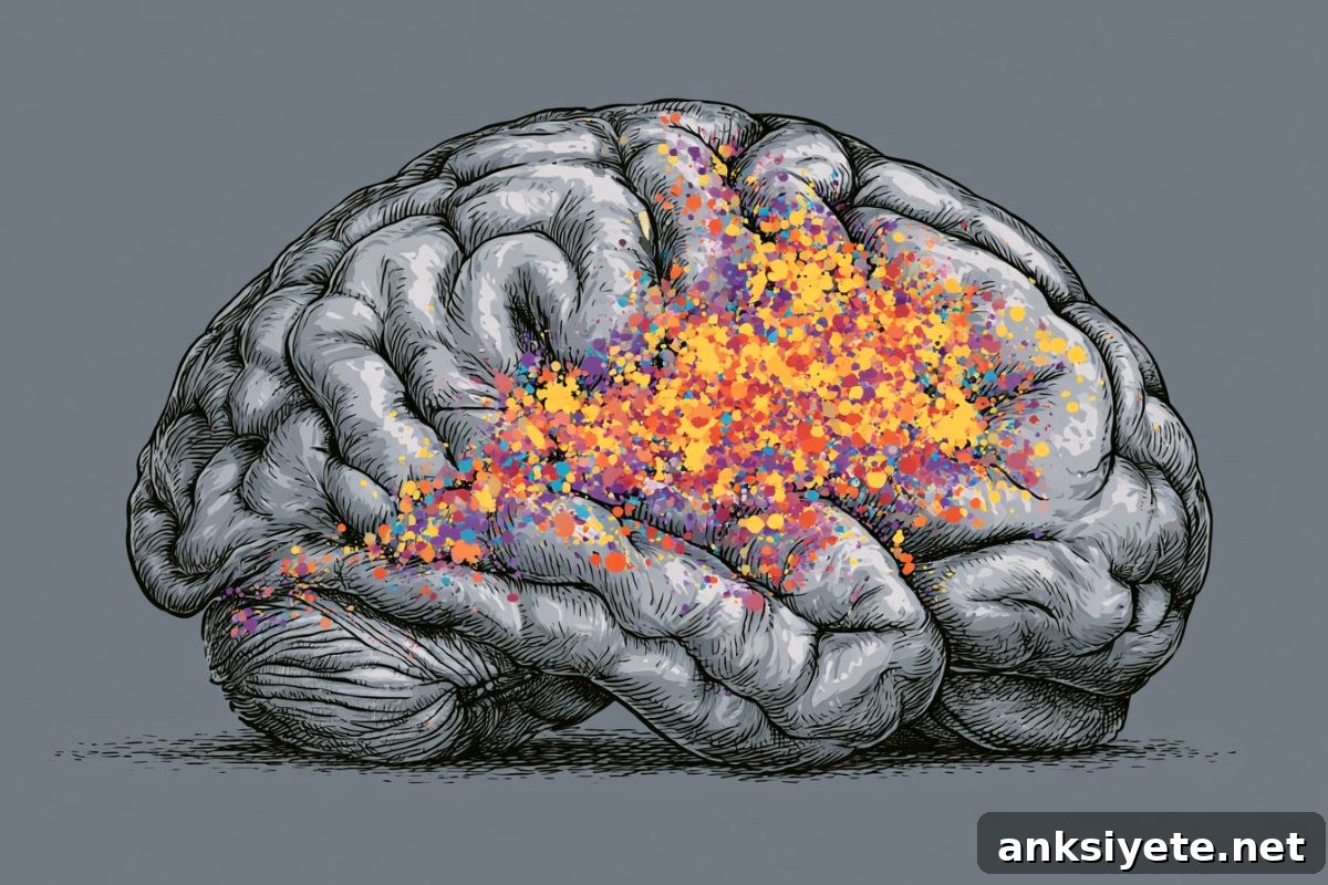
Summary: Why humans and animals need sleep remains one of biology’s longest-standing puzzles. One prominent idea, the synaptic homeostasis hypothesis, suggests that synapses — the connections between neurons — strengthen and multiply during waking hours as we take in and process information. This continuous strengthening consumes energy and generates protein buildup, cluttering neural circuits. Sleep is proposed to serve as a systemic reset that prunes weaker connections and restores balance. While animal studies have mapped this process in detail, direct evidence in humans has been limited.
A new human study supplies important clinical support for this hypothesis. Using advanced positron emission tomography (PET) imaging, researchers measured a molecular marker tied to synaptic density in people who slept normally and in people kept awake for 28 hours. They found measurable, widespread increases in synaptic markers in brain regions responsible for memory and information processing after prolonged wakefulness.
Key Facts
- The metabolic toll of staying awake: Extended wakefulness leads to increased strength and number of synaptic connections in the human brain, raising metabolic demands and supporting the synaptic homeostasis model of sleep.
- SV2A as a structural marker: The study tracked Synaptic Vesicle Glycoprotein 2A (SV2A) using PET scans. SV2A is a widely used biomarker that reflects the density of active synapses in the living brain.
- Specific brain areas affected: After 28 hours without sleep, participants showed elevated SV2A in the hippocampus, a key memory center, and the thalamus, a central sensory and information relay.
- Recovery sleep and slow waves: When sleep-deprived subjects were given a short two-hour nap, those with the highest SV2A increases produced significantly more slow-wave activity, indicating strong sleep pressure and deep recovery sleep.
- Structural changes, not only fatigue: The findings show that sleep loss produces measurable structural changes in neural networks, not merely subjective tiredness.
Source: PLOS
A night without sleep increased markers associated with synaptic connections, suggesting human sleep may be necessary to restore cellular balance in the brain, according to a study published June 23 in the open access journal PLOS Biology by David Elmenhorst and colleagues at the Forschungszentrum Jülich Institute of Neuroscience and Medicine in Germany.
For decades scientists have debated why sleep is essential. One influential explanation is that sleep restores synaptic homeostasis: during wakefulness synapses strengthen as the brain encodes new experiences, raising energy use and producing protein accumulation. Sleep is thought to downscale these changes, pruning synapses and re-establishing equilibrium. Until now, supporting evidence came primarily from animal experiments.
To test this in humans, the research team used PET imaging to measure SV2A in 40 volunteers, half of whom remained awake for a full night. SV2A is present in synaptic vesicles across nearly all central nervous system synapses and serves as a practical proxy for synaptic density in vivo.
After roughly 28 hours of continuous wakefulness, participants in the sleep-deprived group showed higher SV2A signals in multiple brain regions, most notably the hippocampus and thalamus. These increases were modest in magnitude, which aligns with the expectation that the healthy human brain maintains tight homeostatic control to avoid large, potentially harmful shifts in synaptic protein levels.
When the sleep-deprived volunteers were given a two-hour recovery nap, greater SV2A elevations predicted stronger slow-wave activity during that nap. Slow waves are a hallmark of deep sleep and correlate with sleep pressure; their association with SV2A supports the idea that accumulated synaptic strength during wakefulness drives the need for restorative deep sleep.
Although SV2A is an indirect measure of synaptic connectivity and the observed changes were relatively small, the study provides important human evidence consistent with the synaptic homeostasis hypothesis. The researchers conclude that extended wakefulness not only causes subjective fatigue but also produces measurable structural shifts in neural connections that likely contribute to the biological drive to sleep.
Key Questions Answered:
A: SV2A (Synaptic Vesicle Glycoprotein 2A) is a protein located inside the vesicles that store neurotransmitters at synapses. Because SV2A is widespread across synapses in the central nervous system, it acts as a reliable proxy for overall synaptic density. PET imaging of SV2A allows researchers to assess changes in the number or activity of synapses in living human brains as a function of sleep and wakefulness.
A: SHY proposes that wakefulness strengthens synapses as we learn, and sleep downscales those connections to prevent saturation and excessive energy use. By demonstrating that roughly 28 hours of wakefulness increases SV2A levels in specific human brain regions, the study supplies direct human evidence that wakefulness can drive modest increases in synaptic markers, linking accumulated synaptic strength to the biological pressure to sleep.
A: Large, abrupt rises in synaptic structural proteins in a living human brain would be abnormal and potentially pathological. The modest, localized increases observed are consistent with a tightly regulated homeostatic process. Even these small changes were enough to predict how deeply subjects entered slow-wave sleep during a recovery nap, demonstrating that subtle structural shifts can have meaningful functional consequences.
Editorial Notes:
- This article was edited by a Neuroscience News editor.
- The journal paper was reviewed in full.
- Additional context was added by editorial staff.
About this sleep and neuroscience research news
Author: Claire Turner
Source: PLOS
Contact: Claire Turner – PLOS
Image: The image is credited to Neuroscience News
Original Research: Open access. “Learning engages transient and sustained cellular mechanisms in the human brain” by Guillermina Griffa, Marco Palombo, Abraham Yeffal, Hong-Hsi Lee, Agustin Solano, Susie Y. Huang, and Valeria Della-Maggiore. PLOS Biology. DOI: 10.1371/journal.pbio.3003861
Abstract
Learning engages transient and sustained cellular mechanisms in the human brain
Structural neuroplasticity underlies learning and development and influences vulnerability to brain disorders, making it a central focus of neuroscience. Progress in humans has been limited by the difficulty of probing cellular processes in vivo, leaving much of the mechanistic understanding dependent on animal models.
To bridge this gap, researchers combined ultra–high-gradient diffusion MRI with the cell-compartment specificity of the Soma and Neurite Density Imaging (SANDI) model to investigate structural plasticity directly in the living human brain.
Tracking how learning modulates the dynamics of cell bodies and neuronal processes allowed the team to separate plastic from nonplastic microstructural changes. They observed two distinct temporal responses to motor learning: a transient expansion of cell bodies across engaged regions, consistent with a short-lived homeostatic response, and a sustained increase in cell-process density localized to core motor areas, consistent with structural plasticity.
This approach provides a mechanistic view of human neuroplasticity and moves research closer to linking animal findings with human brain function.