
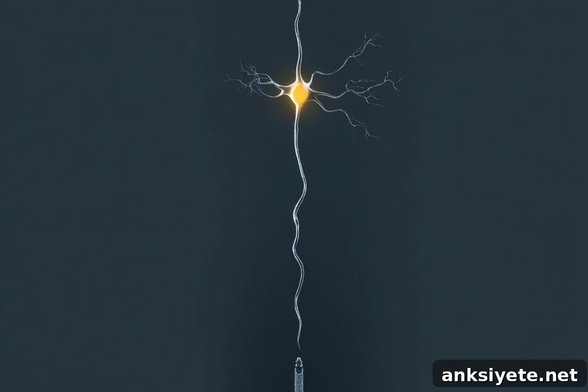
Summary: A major advance in neurotechnology now lets scientists record and control individual neuron activity at depth in the living brain. The new device, Neuropixels Opto, merges large-scale electrophysiological recording with precise optogenetic light delivery on a single silicon shank thinner than a human hair, enabling simultaneous measurement and manipulation of hundreds of cells within intact circuits.
Validated in mice, Neuropixels Opto challenges long-standing assumptions about cortical connectivity and offers a high-resolution experimental platform for mapping circuit dysfunctions linked to disorders such as Alzheimer’s, schizophrenia and Parkinson’s disease.
Key Facts
- One unified neural interface: Neuropixels Opto integrates electrical recording and optogenetic stimulation into a single probe. Previously, researchers had to choose whether to “listen” (electrophysiology) or “control” (optogenetics) because combining both in deep brain tissue introduced electrical noise and technical interference.
- Hair-thin silicon architecture: The probe fits roughly 960–1,000 closely spaced recording sites and an array of microscopic light emitters on a shaft narrower than a human hair. This design captures high-resolution spike data while delivering spatially targeted blue and red light at multiple depths.
- Major international investment: The development is part of a £15 million collaborative initiative supported by the Wellcome Trust, the Allen Institute and partners, led by teams at UCL and the Allen Institute.
- New insight into cortical independence: Experiments using the probe revealed that many cortical neurons can operate with surprising local autonomy rather than always producing large-scale synchronized waves, overturning a common assumption about cortical interconnectivity.
- Direct causal mapping: Because the system can activate or silence selected cell types while recording the same neural population in real time, it enables causal tests of how individual neurons and cell classes drive perception, learning and behavior.
- Applications for brain disorders: By resolving how healthy and diseased circuits differ at cellular resolution, Neuropixels Opto provides a roadmap to develop more targeted diagnostics and interventions for complex neurological and psychiatric conditions.
Source: UCL
Overview
Neuropixels Opto combines two powerful but previously difficult-to-merge approaches: high-density extracellular electrophysiology and optogenetics. Packaged on a single ultra-thin silicon probe, the device records spikes from hundreds of neurons while delivering controlled light pulses to selectively activate or silence genetically identified cell types. Published in Nature Methods, the prototype probes support spatially addressable stimulation with blue and red wavelengths and deliver high-quality recordings in both cortical and deep brain structures.
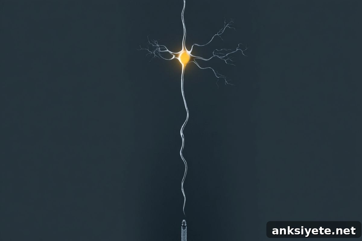
The project was led by visual neuroscience researcher Professor Matteo Carandini and co-lead author Dr Karolina Socha at the UCL Institute of Ophthalmology, in collaboration with teams at the Allen Institute and other international partners. The multidisciplinary effort brought together microfabrication engineers, electrophysiologists and optogenetics specialists to solve the key challenge: delivering light deep into tissue without contaminating sensitive electrical recordings.
At the core of the technology is a 70 µm–wide, centimeter-long silicon shank equipped with around 960 recording pads and multiple columns of embedded light emitters. This combination allows researchers to target distinct depths and locations along the probe for stimulation while capturing detailed spiking activity across the same spatial scale.
In cortex experiments, the probe enabled spatially selective activation and silencing of neurons at different depths and revealed that many cells modulate their activity locally rather than always participating in broad population-wide events. In deeper structures such as the striatum, the system facilitated parallel identification and optotagging of distinct cell types, demonstrating utility for both basic circuit mapping and cell-type-specific investigations.
Implications for neuroscience and medicine
Neuropixels Opto advances the experimental toolkit by converting correlative observations into causal tests: investigators can perturb specific neurons or cell types and immediately observe downstream effects on nearby and distant circuits. This capability will accelerate studies on how precise circuit motifs contribute to perception, decision-making and learning, and it offers a clear path toward identifying circuit-level signatures of neurological disease. The open development model and large-scale collaboration aim to make these tools broadly available to the research community.
Key Questions Answered:
A: Delivering light to control neurons typically introduced electrical noise that swamped sensitive recording electrodes. Traditional setups separated recording and stimulation to avoid interference; Neuropixels Opto overcomes that limitation by integrating photonic and electronic circuits on a single microfabricated probe that minimizes cross-talk.
A: The design uses state-of-the-art micro-silicon engineering to pack roughly 960–1,000 recording sites and two arrays of microscopic light emitters onto a 70-µm-wide shank. Recording pads and emitters are interleaved so the same probe provides both high-resolution sensing and spatially addressable illumination.
A: Experiments showed that many cortical neurons can operate with remarkable locality and independence. Instead of inevitable large-scale activation, selected neurons could be toggled with pinpoint precision while neighboring cells remained relatively unaffected, revising assumptions about cortical synchrony.
Editorial Notes:
- This article was edited by a Neuroscience News editor.
- The journal paper was reviewed in full.
- Additional context was provided by the editorial staff.
About this neurotech research news
Author: Henry Killworth
Source: UCL
Contact: Henry Killworth – UCL
Image: Image credited to Neuroscience News
Original Research: Closed access. “Neuropixels Opto: combining high-resolution electrophysiology and optogenetics” by Anna A. Lakunina et al., published in Nature Methods. DOI: 10.1038/s41592-026-03076-z
Abstract
Neuropixels Opto: combining high-resolution electrophysiology and optogenetics
High-resolution extracellular electrophysiology is a gold standard for recording spikes from distributed neural populations, and it becomes especially powerful when combined with optogenetics for cell-type-specific manipulation with high temporal precision. The Neuropixels Opto probes integrate electronic and photonic circuits, packing approximately 960 recording sites and two sets of 14 light emitters onto a 70-µm-wide, 1-cm-long shank. These devices enable spatially addressable optogenetic stimulation with blue and red light while delivering high-quality recordings. In mouse cortex the probes differentially activated or silenced neurons at distinct depths; in the striatum and other deep structures they enabled efficient optotagging and parallel identification of cell types. Neuropixels Opto probes represent a promising tool for recording, identifying and manipulating neuronal populations at high resolution.