
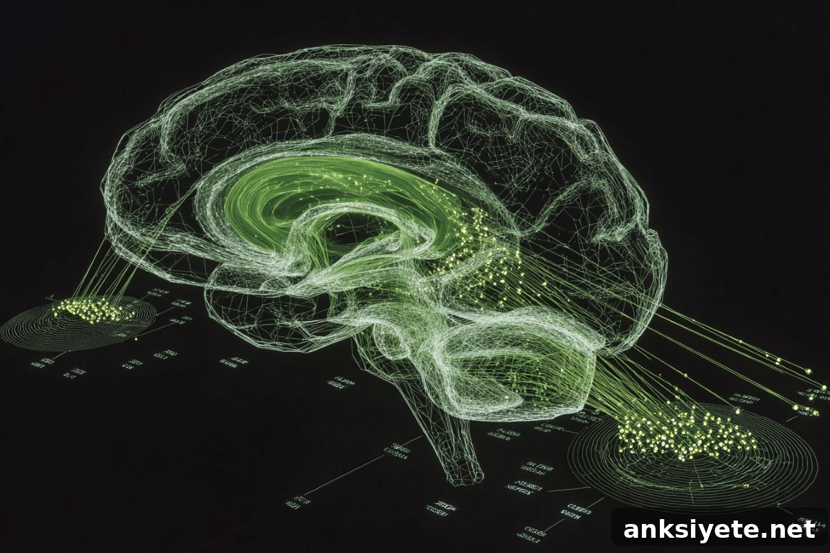
Summary: Researchers overturned decade-old assumptions about how the brain clears metabolic debris by developing a non-disruptive tracking method. They engineered neurons to secrete a traceable fluorescent protein, ZsGreen, and used it to map the exact exit routes for brain-derived proteins.
This approach revealed that proteins do not drain uniformly across the brain. Instead, drainage follows a strict “nearest exit” pattern determined by local anatomy. The study also shows how this system fails in disease: toxic proteins can become trapped inside the brain in Alzheimer’s models or leak directly into the bloodstream during systemic inflammation.
Key Facts
- Limitations of traditional tracers: Injecting dye into cerebrospinal fluid (CSF) has been the standard way to map brain clearance, but it disrupts internal pressures and highlights every potential leak rather than the normal routes used by endogenous proteins.
- ZsGreen neuronal tracing: To avoid these artifacts, researchers engineered mouse neurons to produce a fluorescent reporter, ZsGreen, allowing the team to follow the natural paths that neuron-derived proteins take as they exit the brain.
- Revised drainage map: Contrary to earlier tracer studies that emphasized cervical lymph nodes, most ZsGreen drained through the dura, skull, and nasal cavity, with very little reaching the neck lymph nodes.
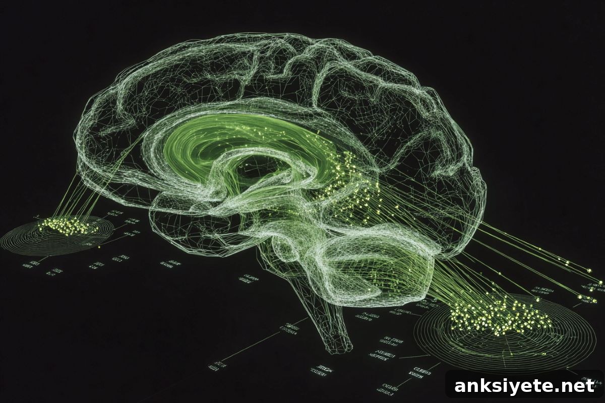
- “Nearest exit” anatomical ZIP codes: The origin of a protein within the brain predicts its exit route: superficial forebrain regions use upper drains, while deep structures like the striatum use routes nearer the skull base. The researchers describe this organization as a biological ZIP code system.
- Immune education via slow outflow: Drainage speed varies by border compartment. Slower clearance at some sites appears to allow border immune cells time to sample neural proteins, helping the immune system learn these molecules as “self” and reduce the risk of autoimmune attack.
- Pathological disruption: Disease alters these routes in different ways. Acute inflammation caused vascular leakage into blood, while Alzheimer’s pathology produced retention inside the brain and obstruction at border exits.
Source: Gladstone Institutes
Analogy: Think of the brain as a well-insulated house with a dedicated plumbing and disposal network. When that infrastructure fails, trash accumulates and the damage is hard to reverse. Similarly, the brain is largely isolated by barriers that tightly regulate exchange with the body, so it relies on specialized clearance systems to remove metabolic waste. Failures in those systems can contribute to neurodegenerative disease.
Historically, researchers introduced external tracers into the CSF to study clearance, but that approach acts much like flooding a house: it reveals all potential leaks without showing which exits endogenous proteins normally use. That left a basic question open—how do proteins made inside the brain find their natural ways out?
Gladstone Institutes researchers developed a genetic, minimally invasive tracing system to answer this question. Described in the journal Cell, the method tracks neuron-produced ZsGreen as it travels from brain tissue to CSF and adjacent border tissues, revealing compartmentalized routes and immune interactions missed by injection-based methods.
Tracking clearance from the source
Instead of introducing dye, the team engineered neurons in mice to secrete ZsGreen and followed its distribution into border compartments such as the dura, skull marrow, nasal cavity, and nearby lymphoid tissue. This approach allowed identification of specific cells at each exit site that interact with neuron-derived proteins—cells that tracer injections had largely overlooked.
The researchers were surprised to find minimal drainage to cervical lymph nodes; most native protein efflux favored dura, skull, and nasal routes. These results highlight the advantage of tracing proteins themselves rather than relying solely on fluid movement as a proxy for clearance.
The “nearest exit” principle
A central discovery was that clearance follows a nearest-exit rule: the anatomical location where a protein is produced largely determines which border it uses to leave the brain. Proteins from upper forebrain areas drain via upper routes, while proteins arising from deeper or basal structures exit through lower pathways. The investigators liken this organization to a ZIP code system that directs waste to the appropriate local exit.
This compartmentalized routing likely contributes to regional vulnerability in disease: when anatomical coordinates or flow patterns change with aging or pathology, waste may accumulate in the wrong locations and promote localized degeneration.
The study also documented differing kinetics across exit sites. Some borders cleared protein rapidly, while others exhibited much slower outflow. Slower clearance may be functionally important because it gives border immune cells time to sample neural proteins and establish tolerance, reducing the risk of autoimmune responses against brain antigens.
Disease insights
Using this tracer system in disease models revealed distinct failure modes. In short-term inflammation, neuron-derived ZsGreen bypassed normal border routes and leaked into the bloodstream. In an Alzheimer’s model, ZsGreen became trapped within brain tissue and failed to exit effectively. These contrasting disruptions point to different therapeutic targets depending on disease context—either stabilizing border compartments to prevent leakage or restoring obstructed exit routes to relieve retention.
Planned follow-up work includes mapping how clearance changes with aging and in other neurological diseases, testing the role of sleep in promoting clearance, and exploring whether tumors manipulate border immune sampling to evade detection.
Funding: This work was supported by the National Institutes of Health (DP5OD033381), the National Institute of Neurological Disorders and Stroke (1R01NS128909, 1RF1NS139975), the Alzheimer’s Association (ADSF-24-1345199-C), the Burroughs Wellcome Fund, the Ludwig Family Foundation, a Longevity Impetus Grant from Norn Group, the UCSF Sandler Program for Breakthrough Biomedical Research, and the Dolby Family.
Key Questions Answered
A: Injected tracers change intracranial pressure and flood the system, highlighting all potential leaks instead of revealing the specific pathways that endogenous proteins use under physiological conditions.
A: The nearest-exit model means the physical origin of a protein within the brain largely determines which border it uses to leave. Each brain region routes its waste to nearby exits, creating a compartmentalized system that functions like a ZIP code to direct clearance.
A: Slower outflow allows border-resident immune cells time to sample neuronal proteins. That exposure helps the immune system learn these molecules as self, reducing the likelihood of damaging autoimmune responses against the central nervous system.
Editorial Notes:
- This article was edited by a Neuroscience News editor.
- The referenced journal paper was reviewed in full.
- Additional context was provided by the editorial staff.
About this neuroscience research news
Author: Julie Langelier
Source: Gladstone Institutes
Contact: Julie Langelier, Gladstone Institutes
Image credit: Neuroscience News
Original Research: Closed access. “Physiological brain clearance architecture revealed by neuronal protein tracing” by Yuichi Chayama, Nalini R. Rao, Daniela Perla, Zimo Zhang, Madigan Reid, Sophia Nelson, Xinlan Wen, Bella Ding, Jessica Blumenfeld, Amanda Apolonio, Sahith Doddipalli, Haoyue Zhou, Sena Gül Turhan, Pu-Yun Shih, Matthias Brendel, Ying-Hui Fu, Ali Ertürk, Zeynep Ilgin Kolabas, Yadong Huang, and Andrew C. Yang. DOI: 10.1016/j.cell.2026.04.048
Abstract
Physiological brain clearance architecture revealed by neuronal protein tracing
Efficient clearance of protein waste is essential for brain homeostasis, yet physiological drainage pathways remain incompletely defined. Traditional tracer injections may not represent endogenous efflux. The authors developed a non-invasive genetic system to trace neuron-derived proteins from the brain to CSF and border tissues. They identify distinct drainage routes and border hotspots missed by injection, confirmed by labeling of endogenous neuronal proteins. Pulse-chase kinetics show slow skull outflow versus rapid dural and nasal clearance. Transcriptomic analyses reveal border cells that sample neuronal antigens, including tolerogenic skull-resident B cells. Regional reporter expression demonstrates compartmentalized clearance consistent with a “nearest exit” principle. Disease models show distinct disruptions: inflammation drives vascular leakage into blood, while amyloid pathology causes parenchymal retention and blocked border exits. These findings describe brain clearance as an organized, compartmentalized network of pathways and immune niches whose dysfunction may underlie regional vulnerability in neurological disease.