
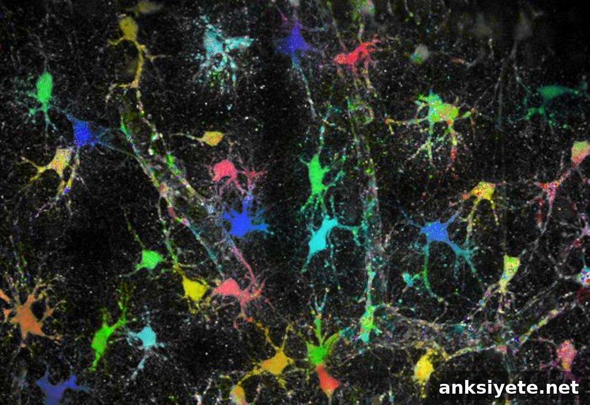
Summary: A new study overturns the long-standing assumption that sodium concentration is uniformly low across astrocytes—the star-shaped glial cells of the brain. Using a novel real-time imaging approach, researchers visualized sodium levels inside astrocytes and their ultra-fine processes for the first time, revealing dynamic, localized sodium micro-domains that adapt to nearby neuronal activity.
Rather than a fixed, uniform baseline, sodium concentrations vary between individual astrocytes and within distinct subdomains of the same cell. These localized differences appear to match the excitability and signaling demands of neighboring neural circuits.
Key Facts
- The Glial Framework: Glial cells—including astrocytes—make up roughly half of the human brain and are essential for brain development, intercellular communication, and maintaining neural network function.
- Sodium and Electrolyte Balance: Positively charged sodium ions (Na+) are critical electrolytes derived mainly from dietary salt. In astrocytes, low intracellular sodium is essential to regulate neurotransmitter clearance at synapses and to keep other ionic balances in check.
- Rethinking Uniformity: Past models assumed a consistent, low sodium baseline across all astrocytes and their fine processes. Direct tissue imaging now shows pronounced heterogeneity in sodium levels both between cells and within cellular compartments.
- Membrane Transport Mechanisms: Collaborative work identified differential expression and arrangement of specific membrane transport proteins—particularly Na+/K+-ATPase (NKA) subunits—that underlie localized sodium handling in astrocyte membranes.
- Multi-Scale Validation: Experimental imaging from Heinrich Heine University Düsseldorf (HHU) informed biophysical simulations at the University of South Florida, and the results were validated in living animal models by teams at the University of Bonn and University Hospital Bonn.
- Clinical Implications: These sodium sub-domains respond dynamically to local synaptic demands. Disruption of such localized ion homeostasis may contribute to neurological conditions where ion regulation fails, including epilepsy and acute stroke, providing new targets for therapeutic research.
Source: HHU
Context and significance
The brain comprises more than just neurons; about half the tissue consists of glial cells, which support development, communication, and the operation of neuronal networks. Astrocytes, a prominent glial subtype with branching, star-like shapes, play a central role in maintaining the chemical environment around synapses.
Sodium ions (Na+) are among the most vital electrolytes in the body, influencing many physiological processes. In the brain, precise control of intracellular sodium in astrocytes is critical for clearing neurotransmitters from synaptic clefts and for balancing other ion concentrations—actions that directly affect neuronal excitability and network stability.
At the Institute of Neurobiology at HHU, Professor Dr. Christine Rose and her team developed an advanced imaging method under the SynGluCross project (funded by the Federal Ministry of Education and Research, BMBF) that makes it possible to measure sodium concentration inside astrocytes and within their microscopic processes in brain tissue. This approach uses multiphoton fluorescence lifetime imaging to quantify sodium within living tissue slices and in vivo.
Partnering with researchers from Friedrich-Alexander-Universität Erlangen-Nuremberg, the University of Bonn, the University Hospital Bonn, and the University of South Florida, the team tested the assumption that astrocytic sodium is uniformly low. Their measurements contradicted that assumption: sodium levels vary significantly across cells and within subcellular domains.
Molecular analyses, including RNAscope and immunohistochemistry, revealed spatial differences in expression of NKA β1 and β2 subunits across astrocytes. Biophysical models incorporating differential NKA expression and variable sodium influx reproduced the experimentally observed heterogeneity, confirming that membrane transport architectures can produce localized sodium micro-domains.
Dr. Jan Meyer, lead author of the study, explained that these specialized sodium sub-domains effectively tune astrocytes to local neural conditions: “We found functionally distinct sub-domains in astrocytes that respond to the immediate needs of nearby neural circuits.”
Professor Christine Rose emphasized the translational potential of the discovery: “These newly identified features of astrocyte physiology may be relevant in disorders where ion homeostasis and neurotransmitter regulation break down, such as epilepsy or stroke. Our findings provide concrete starting points for targeted therapeutic research.”
Key Questions Answered:
A: Astrocytes form highly localized support domains to regulate neurotransmitters and maintain ionic balance. Different synapses and microcircuits have varying activity patterns, so astrocytes create specialized sodium sub-domains in their fine processes that can rapidly adapt to the local demands of neighboring neurons.
A: The discovery began with high-resolution sodium imaging in brain tissue at HHU. Biophysical modelers in South Florida recreated the observed sodium heterogeneity in simulations, confirming the plausibility of the mechanisms. Colleagues in Bonn then validated the patterns in living animal models to demonstrate that the effect is physiologically relevant.
A: These results open a new direction for therapeutic strategies that protect or stabilize the molecular pumps and transporters responsible for local sodium regulation. Since events like seizures and ischemic injury involve catastrophic ion imbalances, targeting the transport mechanisms that maintain sodium micro-domains could help prevent or mitigate neuronal damage.
Editorial Notes:
- This article was edited by a Neuroscience News editor.
- Journal paper reviewed in full.
- Additional contextual information added by staff.
About this neuroscience research news
Author: Arne Claussen
Source: HHU
Contact: Arne Claussen – HHU
Image: Image credit: HHU / Institute of Neurobiology – Jan Meyer
Original Research: Open access. “Cellular and subcellular heterogeneity of astrocytic Na⁺ homeostasis tuning astrocytes into functionally distinct subgroups in the mouse brain” by Jan Meyer, Viola Bornemann, Alok Bhattarai, Sara Eitelmann, Petr Unichenko, Simone Durry, Karl W. Kafitz, Nicholas Chalmers, Jianfeng Fan, Ruth Beckervordersandforth, Christian Henneberger, Ghanim Ullah & Christine R. Rose. Nature Communications. DOI:10.1038/s41467-026-73435-z
Abstract
Cellular and subcellular heterogeneity of astrocytic Na⁺ homeostasis tuning astrocytes into functionally distinct subgroups in the mouse brain
Astrocytes support extracellular ion and neurotransmitter homeostasis, where the inward Na⁺ gradient is a fundamental determinant. Earlier work suggested a uniformly low Na⁺ distribution in astrocytes, implying robust, homogeneous homeostatic properties. Using multiphoton fluorescence lifetime imaging, the authors quantitatively assessed astrocytic [Na+] in mouse brain slices and in vivo and uncovered pronounced cellular and subcellular heterogeneity. These differences correlate with variable capacity for NKA-mediated K+ uptake and are accompanied by distinct spatial expression patterns of NKA β1 and β2 subunits. Biophysical modeling that incorporates differential NKA expression and variable Na+ influx reproduces the experimentally observed heterogeneity. Overall, the results indicate that astrocytes and their subdomains can be functionally specialized by local Na+ homeostasis to meet the needs of surrounding neural networks.