
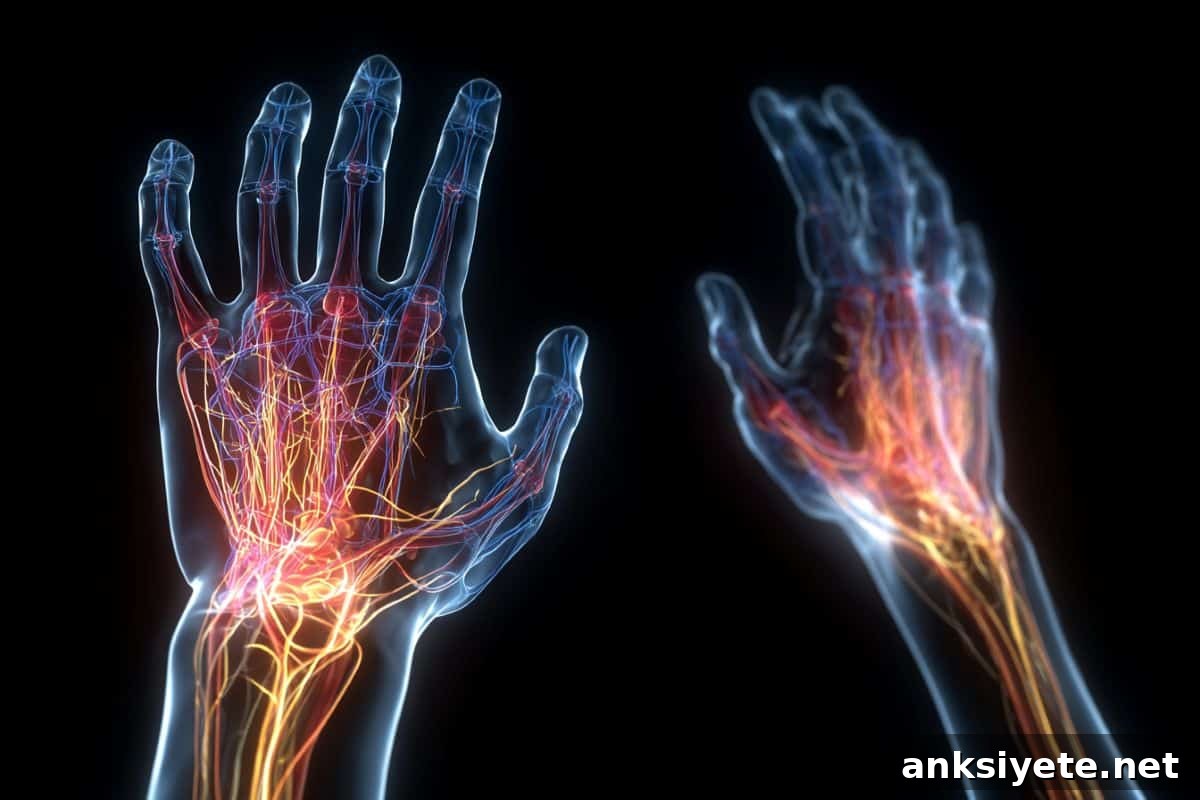
Summary: For decades, neuroscientists assumed that precise, voluntary hand movements were governed almost entirely by the cerebral cortex. New research overturns that idea by identifying an evolutionarily older, conserved network in the brainstem and upper spinal cord that is essential for manual dexterity.
Using comparative fMRI studies in mice and humans, researchers mapped a multi-stage relay system that integrates cortical commands with medullary nuclei and specific cervical spinal segments (C3–C4). This previously underappreciated pathway acts as a complementary route for controlling the hand and could become an important target for therapies aimed at restoring hand function after stroke or other neurological injury.
Key Facts
- New anatomical perspective: Voluntary hand movement travels not only directly from cortex to spinal cord but also through relay centers in the medulla and the C3–C4 propriospinal system in the neck.
- Conserved across mammals: fMRI patterns in mice and humans reveal striking similarities, indicating this circuit is an evolutionarily preserved architecture for limb control.
- Complementary control: The cortex initiates and plans movement, while brainstem and upper spinal circuits coordinate and refine the fine motor details required for grasping and manipulation.
- Clinical potential: Recognizing these alternative pathways offers new targets for neuromodulation or rehabilitation strategies to bypass damaged cortical routes after stroke.
Source: UCR
Researchers led by UC Riverside have uncovered a network linking the brainstem and spinal cord that contributes directly to the control of hand and forelimb movements, revealing an additional layer of neural organization behind our ability to grasp, hold and manipulate objects.
Published in the Proceedings of the National Academy of Sciences, the study demonstrates that voluntary hand commands are routed not only through direct corticospinal projections but also via specific relay centers in the medulla (the lower brainstem) and the top cervical spinal segments. By charting these pathways with functional MRI, the team provides a clearer map of how cortical signals are integrated with older motor circuits to produce precise hand actions.
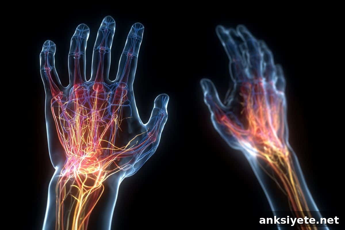
The brainstem sits at the base of the brain and connects to the spinal cord, regulating vital functions such as breathing, posture and balance. The outer cortex is traditionally viewed as the command center for voluntary movement. This study shows that evolutionarily older brainstem regions—the medulla in particular—actively participate in the execution and fine control of hand movements.
“For a long time we believed fine hand control was almost entirely cortical,” said Shahab Vahdat, assistant professor of bioengineering at UCR and lead author of the study. “Our results show that medullary structures and the C3–C4 spinal system also play key roles in shaping these movements.”
The research team identified two medullary regions that consistently activated during hand movement tasks and showed strong functional connectivity with sensorimotor cortical areas. To test this across species, they trained mice to press a small lever with a forepaw while recording brain and brainstem activity and used a similar finger-squeezing task for human volunteers inside the MRI scanner.
Both species showed activation of the same medullary territories and similar patterns of coupling with cortical motor regions, indicating that the corticomedullary network is conserved. Importantly, the study also presents the first human fMRI evidence that the C3–C4 cervical spinal cord segments act as an intermediary, relaying signals from the brainstem to the lower spinal circuits that directly activate hand muscles.
Together, these findings support a multi-stage pathway model: cortical commands are integrated with brainstem nuclei and intersegmental spinal networks before the final motor output reaches the hand. This layered organization helps explain how humans achieve both strength and the fine manipulation required for complex tasks.
Clinically, the discovery has clear implications. Stroke and other injuries that damage cortical motor regions often leave patients with persistent hand deficits. Recognizing alternative, surviving pathways in the brainstem and upper cervical spinal cord opens new avenues for targeted neuromodulation or rehabilitative approaches aimed at recruiting these circuits to restore function.
“These pathways give us additional targets to explore,” Vahdat added. “If therapies can engage or strengthen these routes after cortical injury, they could help compensate for lost function and improve hand and arm recovery.”
Key Questions Answered:
A: Historically, yes — the motor cortex was seen as the principal controller. The new evidence shows the cortex provides high-level planning, while an auxiliary pathway through the medulla and the C3–C4 spinal segments helps implement fine adjustments. Hand control is therefore a multi-stage process rather than a single direct line.
A: Finding the same circuit in mice and humans indicates the mechanism is evolutionarily conserved and fundamental to mammalian limb control. This conservation makes rodents valuable models for mechanistic studies and for developing treatments that could translate to people.
A: When a stroke destroys cortical motor pathways, alternative relays in the brainstem and upper spinal cord may remain intact. Therapies that stimulate or retrain those circuits could help patients recover hand and arm function by rerouting control through these preserved networks.
Editorial Notes:
- This article was edited by a Neuroscience News editor.
- Journal paper reviewed in full.
- Additional context added by editorial staff.
About this motor control and neuroscience research news
Author: Jules Bernstein
Source: UCR
Contact: Jules Bernstein – UCR
Image: The image is credited to Neuroscience News
Original Research: Closed access. “Medullary and C3–C4 propriospinal pathways underlying mammalian forelimb movement control” by Vishwas Jindal, Matteo M. Grudny, Daniel W. Wesson, David E. Vaillancourt, and Shahabeddin Vahdat. PNAS. DOI: 10.1073/pnas.2518217123
Abstract
Medullary and C3–C4 propriospinal pathways underlying mammalian forelimb movement control
Traditional models link goal-directed upper limb actions to cortical motor areas while associating gross postural control with the brainstem. Recent rodent work, however, suggests that medullary nuclei and local spinal circuits also contribute directly to forelimb execution. Whether these findings extend to humans and how medullary, spinal and cortical networks interact dynamically during hand movements remained unclear.
Using fMRI in both humans and mice during forelimb tasks, the study identifies topographically organized corticomedullary networks that include lateral rostral medulla (Lat-RM) and caudal medulla (CauM). In mice, corticomedullary coupling increased along a ventro-medio-dorsal gradient, with the strongest links to primary motor and premotor cortices. In humans, higher-order sensorimotor areas showed the most robust connectivity with CauM and Lat-RM, while a medial rostral medullary region was less engaged.
Simultaneous brain–spinal fMRI in humans revealed distinct functional territories within the C3–C4 cervical cord: ventral zones connected strongly to the medulla while dorsal areas linked more to lower cervical segments. Overall, the findings describe a conserved corticomedullary network that supports forelimb control across species and reveal an indirect pathway—via reticulospinal and C3–C4 propriospinal systems—that contributes to fine hand motor control in mammals.