
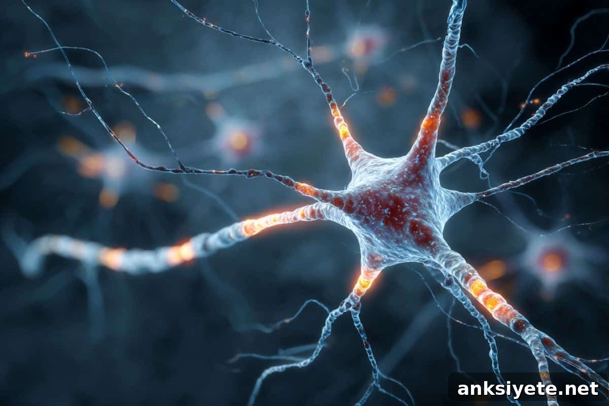
Summary: Researchers have inched closer to explaining why some neurons die while others remain intact in neurodegenerative diseases. By profiling the molecular signatures of neurons from about 80 donors, they identified five distinct neuron subtypes in the motor cortex that are especially vulnerable to the protein TDP-43—implicating these excitatory cells as primary targets in ALS and frontotemporal dementia (FTD).
The study shows that pathological aggregates of the TDP-43 protein largely accumulate in excitatory neurons—cells that transmit and amplify signals in the cortex. This pattern of selective vulnerability indicates that effective therapies will likely need to be directed at these specific high‑risk cell types rather than applied broadly to the entire brain.
Key Facts
- TDP-43 aggregation: Both ALS and FTD commonly feature abnormal clumping of the protein TDP-43 inside neurons, which disrupts cellular processes and contributes to neuron loss.
- Excitatory neurons targeted: The motor cortex excitatory neurons—central to movement control—are disproportionately affected by TDP-43 pathology compared with inhibitory neurons.
- Five vulnerable subgroups: Single‑cell and multi‑omic analyses reveal five distinct excitatory neuron subtypes in the motor cortex that show cell-type specific responses to TDP-43 aggregation.
- Selective vulnerability explained: The molecular mapping helps explain clinical features such as muscle wasting and paralysis: the disease preferentially destroys the cortical neurons that amplify and transmit motor commands.
Source: DZNE
Amyotrophic lateral sclerosis (ALS) and frontotemporal dementia (FTD) are related neurodegenerative disorders that can cause muscle wasting, paralysis, cognitive decline and other severe impairments. At present, effective disease‑modifying treatments are lacking. A shared pathological hallmark in many patients is mislocalization and aggregation of the RNA‑binding protein TDP-43 within neurons.
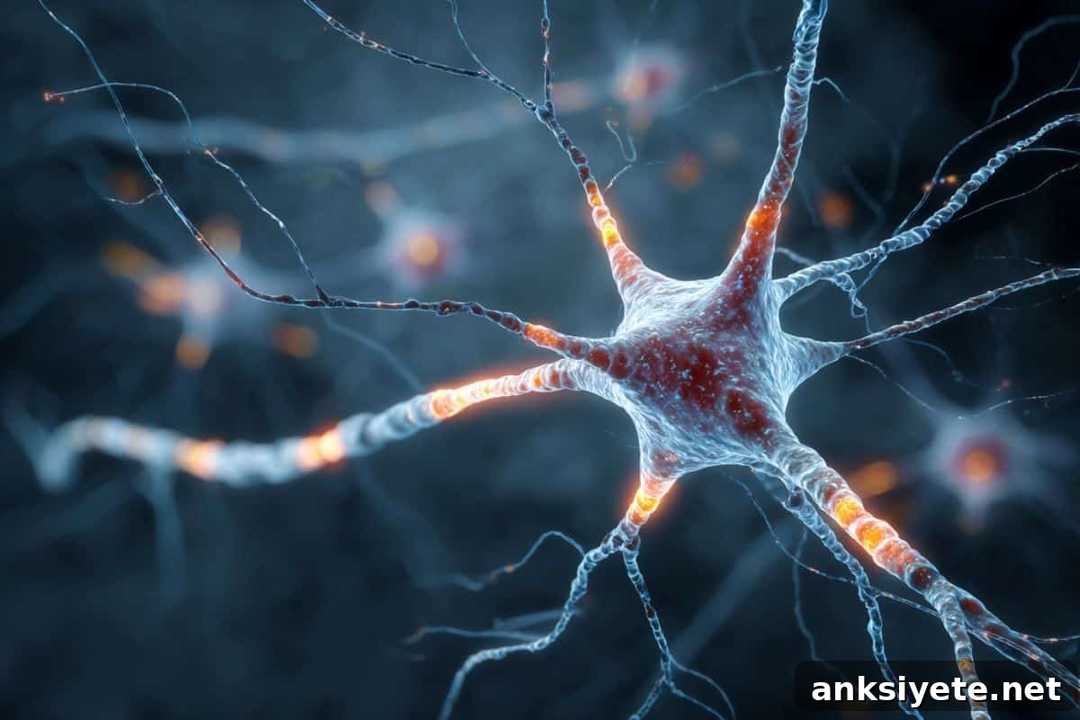
Scientists at DZNE and Ulm University Hospital, collaborating with international partners, examined post‑mortem motor cortex tissue from individuals with ALS, those with a mixed ALS‑FTD diagnosis, and neurologically normal controls. Using advanced sequencing and spatial transcriptomics, the team analyzed neuronal transcriptomes to determine which cell types were affected and how their gene expression profiles shifted in disease.
The motor cortex was selected because it is directly involved in voluntary movement and is therefore central to the motor symptoms of ALS. Across roughly 80 donor samples from Germany, the Netherlands, Scotland and the United States, the investigators found that TDP-43 pathology does not affect all neurons equally. Instead, pathology concentrates in excitatory neurons—those that propagate and amplify cortical signals—pointing to a precision pattern of injury within the motor circuit.
Within the vulnerable excitatory population, five molecularly defined subgroups emerged. Each subgroup displays a distinct transcriptional response to TDP-43 pathology, with specific genes and pathways altered in a subtype‑dependent manner. These differences suggest that different neuron classes experience unique pathological processes and therefore may require tailored protective strategies.
Disease mechanisms
The core of the study is the transcriptome—the complete set of RNA transcripts that reflects which genes are actively expressed in a cell. By comparing transcriptomes from affected versus unaffected neurons, researchers identified cell type‑specific transcriptional abnormalities associated with TDP-43 aggregation, such as altered splicing and cryptic exon inclusion. These molecular signatures clarify how TDP-43 pathology disrupts gene regulation differently across neuron subtypes.
Because the disease signature varies by cell type, the findings argue for therapies that are targeted to the specific vulnerable populations in the motor cortex. Rather than a one‑size‑fits‑all treatment for the whole brain, the data support precision approaches aimed at stabilizing the critical genes and pathways that fail in those five high‑risk neuron subtypes.
Key Questions Answered:
A: It comes down to both location and cell type. The study finds that TDP-43 pathology selectively attacks excitatory neurons in the motor cortex—the brain region that sends commands to muscles. Destruction of these amplifying neurons prevents motor signals from reaching the spinal cord and muscles, producing the movement deficits characteristic of ALS.
A: The molecular fingerprint refers to the transcriptome—the pattern of genes that are turned on or off in a cell. Comparing transcriptomes from healthy and diseased neurons reveals which genetic programs break down in the vulnerable subtypes, effectively mapping the cellular processes disrupted by TDP-43.
A: The findings provide detailed cellular targets and pathways to guide future therapies, but they do not by themselves constitute a cure. They do, however, offer actionable “coordinates” for developing precision treatments that protect or restore the specific neuron subtypes most at risk of TDP-43–related damage.
Editorial Notes:
- This article was edited by a Neuroscience News editor.
- The full journal paper was reviewed.
- Additional context was added by the editorial staff.
About this neurology and ALS research news
Author: Marcus Neitzert
Source: DZNE
Contact: Marcus Neitzert – DZNE
Image: The image is credited to Neuroscience News
Original Research: Open access. “Multi-modal dissection of cell-type specific TDP-43 pathology in the motor cortex” by Wolfgang P. Ruf, Julia K. Kühlwein, Laura Meier, Sarah J. Brockmann, Jaehyun LeeBae, Ghazaleh Sadri-Vakili, Deniz Yilmazer-Hanke, Susanne Petri, Dietmar R. Thal, Veselin Grozdanov & Karin M. Danzer. Nature Communications. DOI: 10.1038/s41467-026-69944-6
Abstract
Multi-modal dissection of cell-type specific TDP-43 pathology in the motor cortex
Cytoplasmic TDP-43 pathology is a central pathological feature of ALS and ALS‑FTD and occurs across a range of genotypes, clinical presentations and central nervous system regions. To develop targeted interventions, it is essential to identify which cell types are affected and what cell-type specific changes underlie their vulnerability.
The study combined flow‑cytometry nuclear sorting with single‑nucleus multi‑omic ATAC‑seq and RNA‑seq as well as spatial transcriptomics to define the transcriptional identity of affected neurons in post‑mortem ALS/ALS‑FTD motor cortex samples (30 ALS, 20 ALS‑FTD and 32 controls).
Results show that TDP‑43 pathology predominantly impacts excitatory cortical neurons and pinpoint the neuron classes most affected: intratelencephalic L2‑L3‑LINC00507‑FREM3, L3‑L5‑RORB‑LNX2, L3‑L5‑RORB‑ADGRL4 and L6‑THEMIS‑LINC00343 neurons, as well as extratelencephalic L5‑FEZF2‑NTNG1 neurons. The transcriptional disturbances caused by TDP‑43, including cryptic exon inclusion and other splicing defects, are distinct for each cell type, underscoring the need for cell‑type specific therapeutic strategies to address TDP‑43 pathology.