
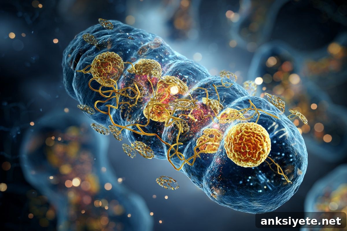
Summary: When the cell’s energy generators—mitochondria—fail, tissues can degenerate, a process central to disorders such as Parkinson’s and Alzheimer’s disease. A recent study from Tokyo University of Science clarifies how healthy mitochondria can be transferred into recipient cells, how cells internalize them, and how those organelles immediately restore bioenergetic function.
Investigators showed that mesenchymal stromal cells (MSCs) actively take up isolated, functional mitochondria through several endocytic pathways. Once internalized, these mitochondria rapidly increase ATP production, improve oxidative stress resistance, and accelerate cellular repair and proliferation. The findings strengthen the scientific foundation for mitochondrial transplantation — an emerging therapeutic approach that may be applied to acute injuries and chronic diseases driven by mitochondrial dysfunction.
Key Facts
- Deliverable energy units: Functional mitochondria can be isolated from healthy cells and delivered to energy-deficient cells, restoring ATP production and respiratory capacity.
- Active cellular uptake: Recipient MSCs internalize mitochondria using multiple endocytic routes, including clathrin-dependent, caveolin-associated, and actin-mediated mechanisms.
- Rapid functional gains: Internalized mitochondria increase cellular respiration, boost antioxidant defenses, and promote proliferation in a concentration-dependent manner.
- Non-genetic approach: Mitochondrial transplantation supplements cellular bioenergetics without altering DNA, offering a potentially faster and safer option than gene-based therapies for acute conditions.
- Therapeutic potential: This strategy could be relevant for ischemia–reperfusion injuries, toxin-induced organ damage, and neurodegenerative diseases where mitochondrial failure is central.
Source: Tokyo University of Science
Why mitochondria matter
Mitochondria produce the majority of a cell’s usable chemical energy as adenosine triphosphate (ATP). Beyond energy production, they regulate programmed cell death, calcium balance, and cellular responses to stress. Loss of mitochondrial function undermines a cell’s ability to meet energetic demands and maintain homeostasis, which contributes to neurodegeneration, inflammatory conditions, and metabolic disorders.
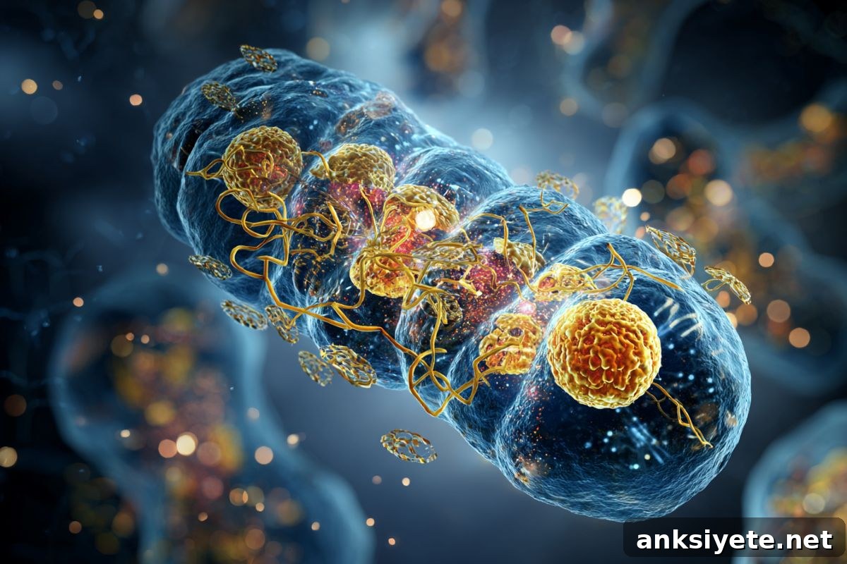
Scientists are actively investigating therapies that restore mitochondrial function directly. Mitochondrial transplantation is an approach in which functional mitochondria are isolated from donor cells and delivered to compromised tissues to replenish energy supply and support recovery.
Progress in this field has been limited by a fundamental gap: the mechanisms by which recipient cells interact with and internalize isolated mitochondria were not fully understood. Do mitochondria act extracellularly, or must they enter the cell to be effective? If uptake occurs, which cellular pathways mediate it, and do the organelles remain functional inside recipient cells?
To answer these questions, a team led by Associate Professor Kosuke Kusamori at Tokyo University of Science used mesenchymal stromal cells (MSCs) as a model system. Combining biochemical assays with advanced imaging — fluorescence and confocal microscopy, label-free live imaging, flow cytometry, and electron microscopy — the researchers examined mitochondrial isolation, uptake dynamics, and post-uptake function.
They isolated mitochondria with methods that preserved structural integrity and confirmed the preparations were free of contaminating cellular components. Isolated mitochondria retained the capacity to produce ATP. When supplied to living MSCs and hepatocytes, these mitochondria increased proliferation, improved survival under chemical and oxidative stress, and elevated oxygen consumption rates, consistent with enhanced bioenergetic function.
Time-course imaging and flow cytometry showed progressive internalization of mitochondria over hours, with electron microscopy revealing mitochondria-like structures inside membrane-bound vesicles within recipient cells. By inhibiting specific endocytic pathways pharmacologically, the team demonstrated that MSCs rely on multiple, overlapping endocytic mechanisms rather than a single dominant route to take up mitochondria.
These observations provide direct evidence that isolated mitochondria are actively incorporated into MSCs and retain biological activity after internalization. The results help explain how mitochondrial transplantation can produce rapid cytoprotective and proliferative effects in donor cells.
Dr. Kusamori comments that this work lays the groundwork for a new medical field — mitochondrial therapy — which aims to restore cellular energy supply as a therapeutic strategy. Because mitochondrial transplantation does not alter nuclear DNA, it could be positioned alongside stem cell and gene therapies as a complementary, non-genetic approach, particularly suited to acute or localized mitochondrial dysfunction.
Despite promise, translation to clinical practice requires additional research: optimizing delivery methods for different tissues, controlling uptake and distribution, ensuring product purity and functional integrity, assessing long-term safety and efficacy, and preventing unwanted immune responses. Mitochondrial transplantation remains preclinical and will need extensive validation in disease models before human trials begin.
Key Questions Answered:
A: Conceptually yes. Mitochondrial therapy involves delivering healthy, functional mitochondria to cells with compromised bioenergetics. This study demonstrates that recipient cells can internalize donated mitochondria and begin using them to restore energy production.
A: Many current treatments focus on symptoms. Mitochondrial transplantation aims to address an underlying cause in many neurodegenerative conditions—the loss of cellular energy—by restoring ATP production and improving cellular resilience, which could slow degeneration if successfully translated to patients.
A: Because it uses naturally occurring organelles and does not alter nuclear DNA, it is considered a potentially safer alternative to genetic modification. However, safety must be established in preclinical studies and clinical trials to rule out immune reactions, off-target effects, or other risks.
Editorial Notes:
- This article was edited by a Neuroscience News editor.
- The original journal paper was reviewed in full by the editorial team.
- Additional context and clarifications were added by staff to aid understanding.
About this neurology research news
Author: Yoshimasa Iwasaki ([email protected])
Source: Tokyo University of Science
Contact: Yoshimasa Iwasaki – Tokyo University of Science
Image: Image credited to Neuroscience News
Original Research: Uptake mechanisms and functions of isolated mitochondria in mesenchymal stromal cells. Mai Kanai, Miyabi Goto, Shoko Itakura, Makiya Nishikawa & Kosuke Kusamori. Scientific Reports. DOI: 10.1038/s41598-025-28494-5.
Abstract
Uptake mechanisms and functions of isolated mitochondria in mesenchymal stromal cells
Mitochondrial transplantation is a promising therapeutic strategy, but mechanisms of interaction and internalization by recipient cells were unclear. This study isolated functional mitochondria from mesenchymal stromal cells and characterized their bioenergetic activity and physicochemical properties. Treatment with isolated mitochondria promoted cell proliferation, improved survival under stress, and increased oxygen consumption rates, indicating enhanced cellular bioenergetics. Time-course imaging and quantitative assays showed progressive mitochondrial internalization within 24 hours. Chemical inhibition of endocytosis revealed that multiple endocytic pathways contribute to mitochondrial uptake. The findings indicate that MSCs actively incorporate isolated mitochondria and that internalized organelles retain functional activity, supporting the potential application of mitochondrial therapy in various disease contexts.