
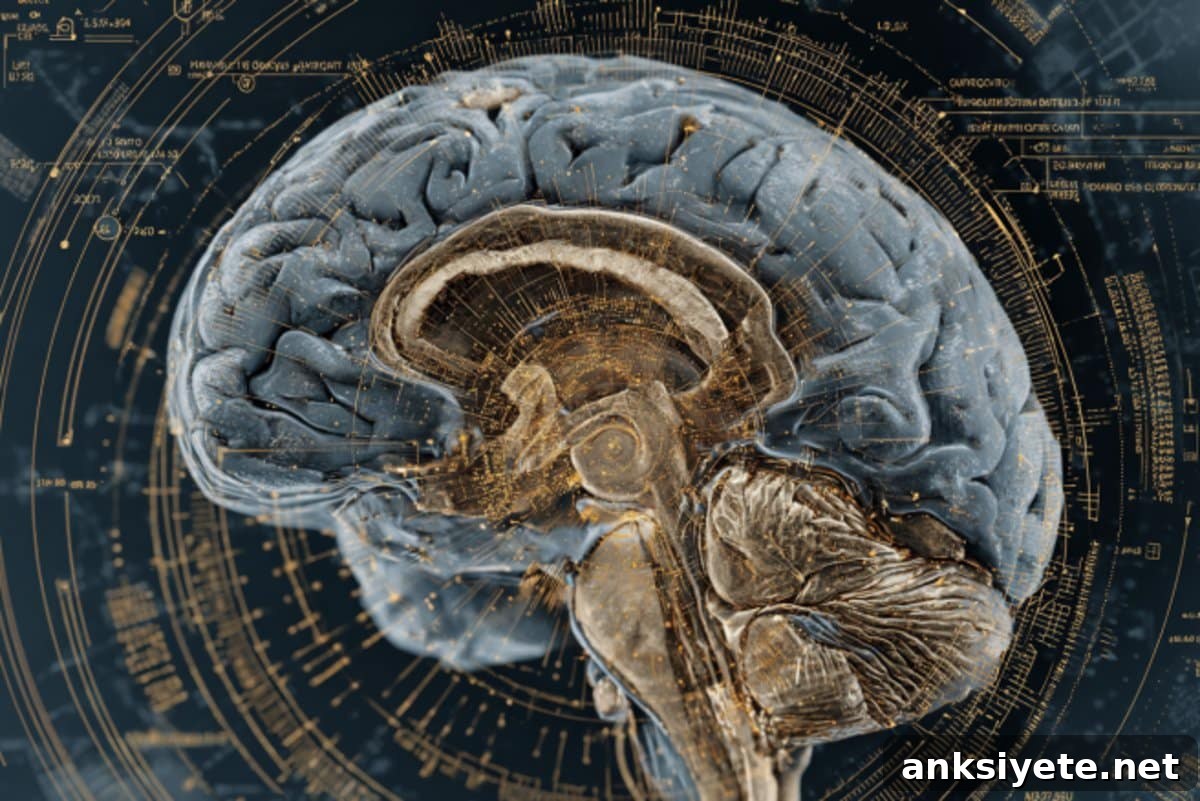
Summary: NextBrain is a new AI-powered, histology-based brain atlas that lets researchers map the adult human brain at an unprecedented level of anatomical detail. Built by aligning roughly 10,000 microscopic tissue sections from post-mortem brains with MRI using advanced AI, the atlas provides a 3D, probabilistic map of 333 precise brain regions that were previously invisible on standard MRI scans.
When applied to thousands of in vivo and ex vivo MRI scans, NextBrain reliably identified fine-grained structures faster and more consistently than prior tools. The freely available atlas is intended to accelerate research into brain development, ageing and neurodegenerative conditions, improving opportunities for earlier diagnosis and the development of targeted treatments.
Key Facts
- High precision: NextBrain delineates 333 regions across the adult human brain, enabling subregional analysis that approaches cellular-level detail when inferred from MRI.
- AI-enabled integration: Artificial intelligence aligned histological microscopy and MRI from approximately 10,000 tissue slices to create a coherent three-dimensional atlas.
- Clinical relevance: The atlas makes it possible to detect subtle structural changes associated with conditions such as Alzheimer’s disease, supporting earlier and more specific assessments.
Source: UCL
Overview
The human brain is made up of hundreds of interconnected regions. Conventional MRI atlases identify major structures—like the hippocampus, important for memory—but they lack consistent labels for many smaller subregions. Those subregions can be differently affected by ageing and disease, so distinguishing them matters for both research and clinical practice.
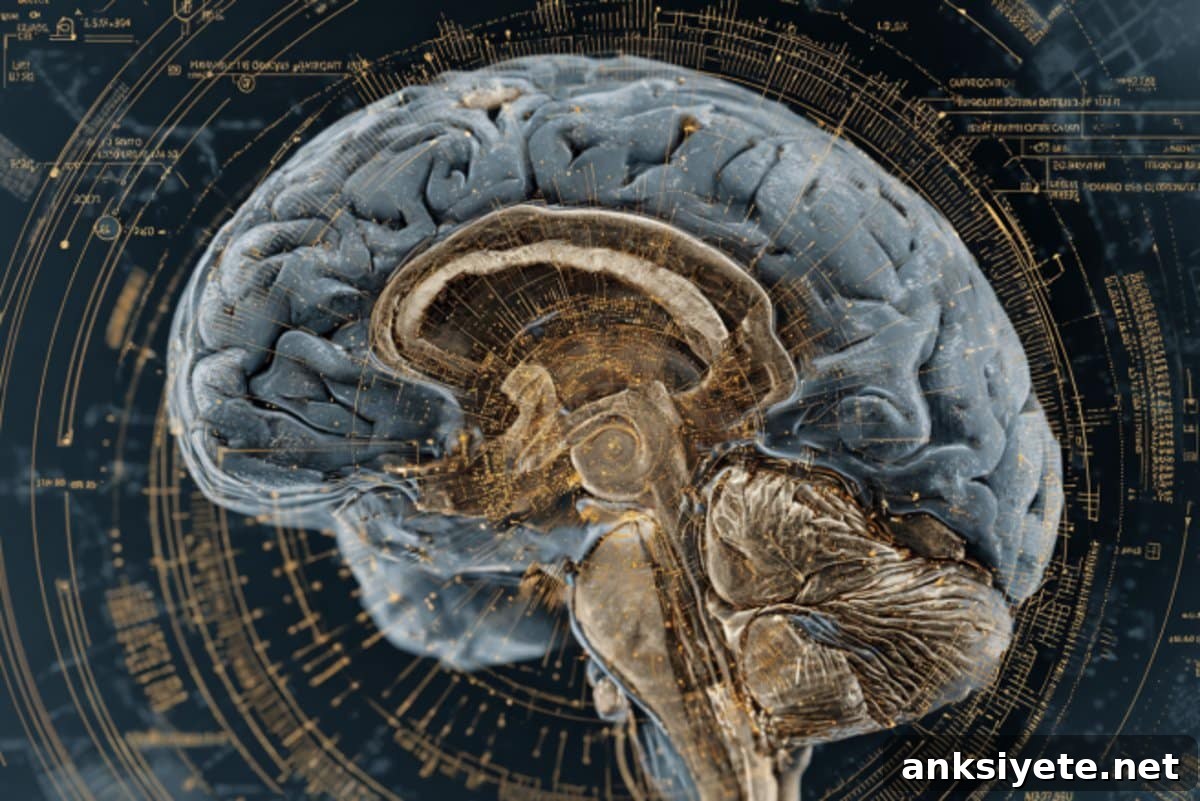
Microscopy (histology) reveals cellular architecture but requires post-mortem tissue, so it cannot be used directly for living subjects. NextBrain bridges that gap by using AI to transfer histological detail into an MRI-compatible 3D atlas that can be applied automatically to scans of living people.
Published in Nature, NextBrain represents a major advance in neuroimaging. The research team built a probabilistic, whole-brain histological atlas and released the resources openly so researchers worldwide can apply it to their MRI studies.
How NextBrain was created
Developing the atlas was a meticulous, multi-year project involving five adult human brains. Each brain hemisphere was sectioned into thousands of thin slices (around 10,000 total), stained to reveal cellular features, and photographed under a microscope. Before sectioning, each brain was scanned with MRI so the team could later reassemble the slices into an anatomically consistent 3D model.
AI tools were essential for aligning the microscope images with the MRI volumes, compensating for distortions introduced during tissue processing and ensuring accurate, gap-free reconstruction. AI also accelerated the labelling process: the team delineated 333 regions across the five reconstructed brains, a task that would have taken decades if done entirely by hand.
The final atlas is an averaged, probabilistic model of those five brains, designed to generalize to the adult population. It can be used to infer high-resolution anatomical detail from routine MRI scans of living or post-mortem subjects.
Validation and applications
The team validated NextBrain on thousands of MRI datasets collected with different scanners and protocols. In one test, the atlas automatically labelled regions in an ultra-high-resolution ex vivo MRI; these automated labels closely matched manual delineations, including small hippocampal subregions. In another large study, NextBrain was applied to over 3,000 in vivo MRI scans to study age-related volume changes, producing finer-grained and more anatomically specific results than previously possible.
Researchers involved in the project highlight the atlas’s potential to enable rapid, reproducible identification of hundreds of brain regions in clinical and research MRI. By bringing microscopic anatomical accuracy into routine imaging, NextBrain opens new opportunities to detect early pathological changes—such as those that occur in Alzheimer’s disease—well before clinical symptoms appear.
A: NextBrain is an AI-assisted, probabilistic histological atlas of the adult human brain that enables high-resolution segmentation of MRI scans, improving the anatomical specificity of neuroimaging research and clinical assessments.
A: The atlas was built by registering roughly 10,000 histological sections from five post-mortem hemispheres to pre-sectioning MRI scans, using AI to align images and label 333 regions in three dimensions.
A: NextBrain enables detection of subtle regional brain changes in living individuals, aiding earlier diagnosis, more precise monitoring of neurodegenerative diseases, and deeper study of brain ageing and development.
About this research
Author: Matt Midgley
Source: UCL
Contact: Matt Midgley – UCL
Image credit: Neuroscience News
Original Research: Open access. “A probabilistic histological atlas of the human brain for MRI segmentation” by Juan Eugenio Iglesias et al., published in Nature.
Research abstract (summary)
Brain atlases are fundamental to neuroimaging because they enable segmentation of regions of interest and comparisons across subjects. Existing histology-derived atlases provide detailed cytoarchitectural maps but often lack probabilistic labels across the whole brain. NextBrain fills this gap by offering a whole-brain probabilistic histological atlas.
The authors developed AI-enabled pipelines to align about 10,000 histological sections from five hemispheres into three-dimensional volumes and to delineate 333 regions of interest. A companion Bayesian segmentation tool allows automated assignment of these regions in MRI scans. The atlas was validated on ultra-high-resolution ex vivo MRI and on large-scale in vivo MRI studies of ageing and Alzheimer’s disease.
By making raw and aligned data, visualization tools, the atlas itself, segmentation software, and ground-truth delineations publicly available, the project aims to democratize access to high-resolution anatomical maps and accelerate discoveries about the healthy and diseased human brain.
Funding: The work received support from the European Research Council, Alzheimer’s Society, the Lundbeck Foundation, and the U.S. National Institutes of Health.