
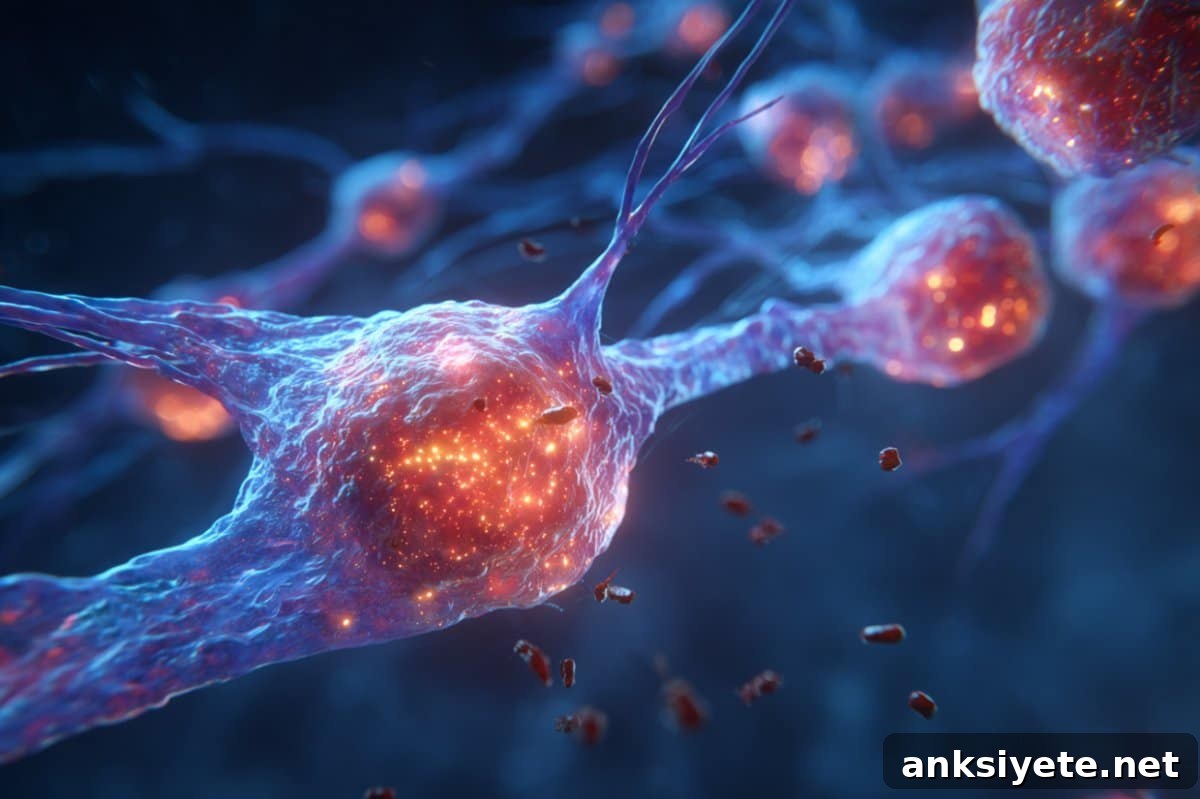
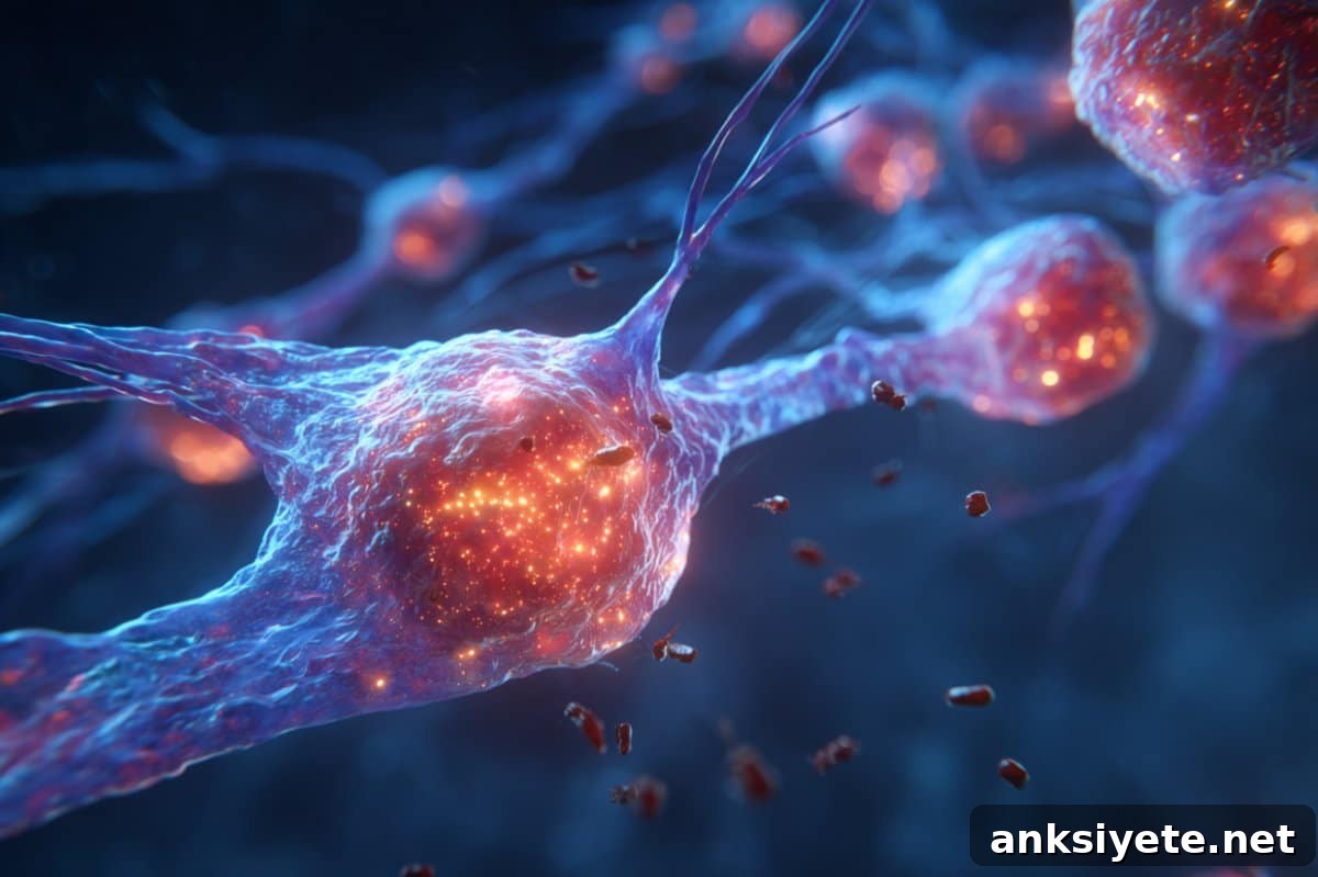
Summary: The retina mounts a distinct immune response when photoreceptor cells are damaged: instead of recruiting circulating neutrophils—the immune system’s typical first responders—the retina relies on microglia, its resident immune cells. This localized response appears to limit inflammation and protect delicate retinal tissue from further harm.
Researchers using advanced adaptive optics imaging observed these dynamics in live mouse retinas. Their results point to a retina-specific mechanism that “cloaks” injury sites and prevents the influx of systemic immune cells, a finding that could inform new approaches to treating diseases that cause vision loss.
Key Facts:
- Retina-specific immune behavior: Neutrophils—despite being present in nearby blood vessels—are not recruited to repair damaged photoreceptors.
- Microglia lead the response: Brain- and retina-resident microglia migrate to injury sites and handle early repair activities.
- Protective inflammation control: The retina appears to suppress broad immune cell recruitment, likely to avoid collateral damage from excessive inflammation.
Source: University of Rochester
Background: In most tissues, circulating neutrophils rush to sites of infection or injury as a rapid defense. The retina, however, behaves differently. Scientists at the Flaum Eye Institute and the Del Monte Institute for Neuroscience at the University of Rochester found that when photoreceptor cells are injured, the retina calls on microglia rather than neutrophils, even when neutrophils are traveling in vessels mere microns away.
“This discovery has broad implications for the millions of people affected by photoreceptor loss and vision impairment,” said Jesse Schallek, PhD, associate professor of Ophthalmology and senior author of the study published in eLife. “Understanding how microglia and neutrophils interact—or do not interact—in the retina is critical as we develop therapies that must navigate the immune environment of the eye.”
The team used adaptive optics imaging, a high-resolution camera technology developed at the University of Rochester, to visualize single neurons and immune cells in living mouse eyes. This approach allowed the researchers to observe immune cell behavior in real time as photoreceptor damage unfolded.
Their imaging showed that while both microglia and neutrophils are present in and around the retina, only microglia actively migrate into the site of photoreceptor injury. Neutrophils, although passing through adjacent blood vessels, do not enter the injury locus or contribute to the early repair process. Post-mortem confocal microscopy confirmed the in vivo observations.
The researchers interpret these results as evidence of a protective mechanism that limits recruitment of systemic immune cells. Such a mechanism would reduce the risk of collateral damage from widespread inflammation, which can be especially harmful in the highly specialized retinal tissue where photoreceptors are essential for vision.
“It’s striking that neutrophils travel so close to reactive microglia yet remain uninvolved,” Schallek added. “This contrasts with responses in other tissues, where neutrophils are typically the first to respond and drive a robust inflammatory reaction.”
Photoreceptors are specialized retinal neurons that convert light into electrical and chemical signals, which are then sent to the brain so we can see. Many retinal diseases—including age-related macular degeneration, retinitis pigmentosa, and cone-rod dystrophy—lead to photoreceptor death and currently lack cures. Mapping the immune dynamics that follow photoreceptor injury is therefore important for designing interventions that preserve or restore vision.
This study demonstrates the ability to visualize the behavior of individual immune cells in vivo as they interact during retinal injury. These insights will help researchers and clinicians better predict how therapies—particularly those that engage the immune system—might perform inside the eye.
The study was led by first author Derek Power, a laboratory technician in the Schallek lab. Co-authors include Justin Elstrott, PhD, of Genentech, Inc., among others.
Funding: The research received support from the National Eye Institute, Research to Prevent Blindness, the Dana Foundation, and a Collaborative Research Grant from Genentech, Inc.
About this visual neuroscience research news
Author: Kelsie Smith Hayduk ([email protected])
Source: University of Rochester
Contact: Kelsie Smith Hayduk – University of Rochester
Image credit: Neuroscience News
Original Research: Open access. “Photoreceptor loss does not recruit neutrophils despite strong microglial activation” by Derek Power et al., published in eLife.
Abstract
Photoreceptor loss does not recruit neutrophils despite strong microglial activation
When the central nervous system (CNS) is injured, both tissue-resident immune cells such as microglia and circulating neutrophils often act as first responders. How these two immune populations interact after CNS damage is not well understood, and the neural retina presents particular imaging challenges for resolving these dynamics in vivo.
In this study, the authors used fluorescence adaptive optics scanning light ophthalmoscopy (AOSLO) alongside label-free phase-contrast AOSLO to track microglia and neutrophil behavior in mice at micron-level resolution. Retinal lesions were created by focusing 488 nm light onto photoreceptor outer segments, producing focal photoreceptor ablation with minimal collateral injury above or below the plane of focus.
In vivo AOSLO and optical coherence tomography (OCT) were used to follow the cellular response from minutes to months after injury. Microglia responded dynamically and progressively, migrating into the injury site within one day. Neutrophils, despite being carried in vessels only microns from the lesion, were not recruited to the damaged photoreceptors. Post-mortem confocal imaging corroborated these in vivo findings.
These results indicate that acute, focal loss of photoreceptors triggers strong microglial activation without recruiting neutrophils—highlighting a retina-specific immune response relevant to many retinal diseases.