
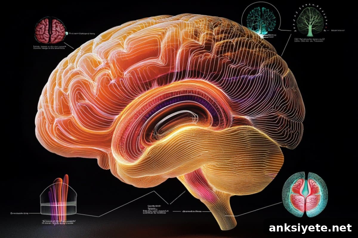
Summary: New research shows that small, shallow grooves on the human brain—known as tertiary sulci—are closely associated with reasoning skills and functional brain connectivity in children and adolescents. These uniquely human folds appear to reduce distances between specific brain regions, improving communication and supporting higher cognitive functions.
In an fMRI study from the University of California, Berkeley, greater depth of tertiary sulci in lateral prefrontal and lateral parietal cortices correlated with stronger functional connectivity and better reasoning performance. The researchers propose that sulcal depth could become a useful biomarker for tracking cognitive development or identifying atypical neurodevelopment.
Key facts:
- Tertiary sulci and cognition: Small, late-developing cortical folds are linked to reasoning ability in youth.
- Improved connectivity: Deeper sulci may physically shorten pathways between regions, supporting faster or more efficient neural signaling.
- Developmental marker: Individual differences in sulcal morphology may help explain variation in cognitive performance and offer diagnostic insight.
Source: UC Berkeley
Many of the grooves and dimples that cover the human cerebral cortex are distinct to our species. Historically they were sometimes dismissed as a byproduct of fitting a large brain into a confined skull, but growing evidence suggests these folds play functional roles. The latest study links the depth of some of the smallest cortical grooves—the tertiary sulci—with increased interregional connectivity and improved reasoning.
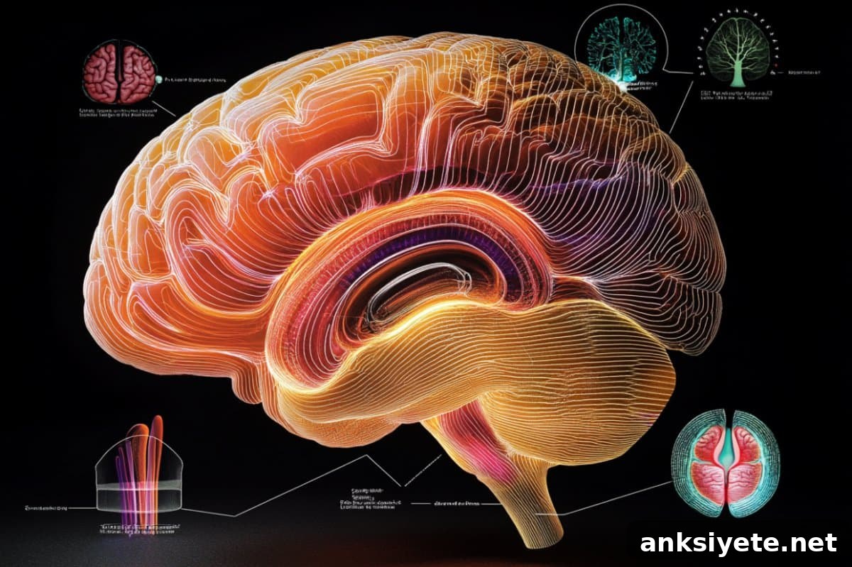
Published May 19 in The Journal of Neuroscience, the study examined 43 participants aged 7–18. The team measured sulcal anatomy and recorded brain activity while participants performed a reasoning task in an fMRI scanner. They focused on sulci in the lateral prefrontal cortex (LPFC) and the lateral parietal cortex (LPC)—regions known to support reasoning, planning and other high-level cognitive functions.
The results indicate that several tertiary sulci involved in reasoning are deeper in individuals whose prefrontal and parietal sulci show higher network centrality. In practical terms, deeper grooves may draw associated regions closer together, shortening the connections between them and potentially increasing neural efficiency during reasoning tasks.
The hills and valleys of the brain
Most non-primate mammals have largely smooth brains, while primates show increasing cortical folding. Humans have especially deep and complex sulci, with an estimated 60–70% of the cortex tucked into these folds. Cortical folding patterns form late in prenatal development and can shift modestly through life, though each person’s overall sulcal configuration—size, shape, location and sometimes even presence or absence of particular sulci—tends to remain a stable individual trait.
Tertiary sulci are the smallest of these grooves. They appear late in prenatal development, remain shallower than primary sulci, and many are unique to humans. Scientists have proposed that tertiary sulci emerge in brain regions that expanded most during human evolution and that their protracted development parallels the long maturation of complex cognitive abilities such as reasoning, decision-making and self-control.
Until recently, direct evidence tying tertiary sulci to brain connectivity and cognition was sparse. This UC Berkeley study adds to a small but growing body of work demonstrating meaningful links between tertiary sulcal anatomy and functional brain networks.
Sulci linked to cognition
The investigators—led by psychologists and neuroscientists at UC Berkeley—have studied how individual differences in sulcal anatomy relate to behavior. One line of earlier work examined the mid-fusiform sulcus in visual cortex and found that its length predicts face-recognition ability; shorter and shallower versions were associated with developmental prosopagnosia in some individuals.
Building on that foundation, the team mapped smaller sulci across lateral prefrontal and lateral parietal cortices. In a previous 2021 study, they developed a model showing that specific tertiary sulci in LPFC accounted for substantial variation in reasoning ability among children. The current study expanded the mapping to LPC and analyzed functional connectivity among 21 sulci per hemisphere, including for the first time detailed analysis of tertiary sulci in these regions.
Across participants, deeper tertiary sulci in several locations were associated with greater network centrality within the prefrontal–parietal sulcal network. In other words, sulcal morphology corresponded with how centrally a sulcus participated in task-related functional networks, providing a plausible anatomical mechanism linking sulcal depth and reasoning performance.
Experience and plasticity
The researchers emphasize that sulcal depth is not a fixed destiny for cognitive ability. Sulcal morphology does not explain all variance in reasoning, and experience—such as educational quality and learning opportunities—strongly shapes cognitive development. The brain remains plastic through childhood, adolescence and even into adulthood, and environmental factors can alter cognitive trajectories.
To support broader research, Weiner’s lab is developing software to automatically identify tertiary sulci. Most existing tools detect roughly 35 sulci, but when tertiary sulci are included the count increases to over 100; the team has also documented new sulci through their work. Using sulci as individualized anatomical landmarks may improve comparisons between brains and reduce mismatches that arise from group-average brain maps.
The authors argue that anchoring functional connectivity analyses to each person’s sulcal anatomy offers a promising way to reconcile diverse brain maps and to extract network-level insights tied to local anatomy.
The study’s co-authors include Silvia Bunge and Kevin Weiner, along with former postdoctoral fellow Suvi Häkkinen, former graduate student Willa Voorhies, former undergraduates Ethan Willbrand and Jewelia Yao, and former visiting scholars Yi-Heng Tsai and Thomas Gagnant.
Funding: This research was supported by the National Institutes of Health (Eunice Kennedy Shriver National Institute of Child Health and Human Development R21HD100858; National Institute of Mental Health R01MH133637) and the National Science Foundation (CAREER Award 2042251).
About this neuroscience and neurodevelopment research news
Author: Robert Sanders
Source: UC Berkeley
Contact: Robert Sanders – UC Berkeley
Image: The image is credited to Neuroscience News
Original research: Closed access. “Anchoring functional connectivity to individual sulcal morphology yields insights in a pediatric study of reasoning” by Silvia Bunge et al., Journal of Neuroscience (DOI: 10.1523/JNEUROSCI.0726-24.2025)
Abstract
Anchoring functional connectivity to individual sulcal morphology yields insights in a pediatric study of reasoning
Cortical folding is a defining feature of the human brain, and mounting evidence links sulcal morphology to functional brain architecture and cognition. Prior work identified that the depths of three small sulci in lateral prefrontal cortex relate to reasoning performance during development. The current study examined the mechanism linking sulcal anatomy and cognition by analysing functional connectivity among sulci in lateral prefrontal and lateral parietal cortices.
Using manual parcellations (21 sulci per hemisphere, 1,806 total) and fMRI from a reasoning task in 43 participants aged 7–18, the authors performed clustering and classification analyses of sulcal-level functional connectivity. Key findings: (1) individual sulci exhibit distinct connectivity patterns; (2) sulcal connectivity does not consistently match probabilistic region labels or large-scale network templates; (3) sulci cluster by shared connectivity profiles rather than mere spatial proximity; and (4) greater depth in several sulci corresponds with higher network centrality. These results support the idea that sulcal morphology is relevant for functional connectivity and suggest that using sulci to define individualized coordinate spaces for functional connectomes is a promising direction for future research.