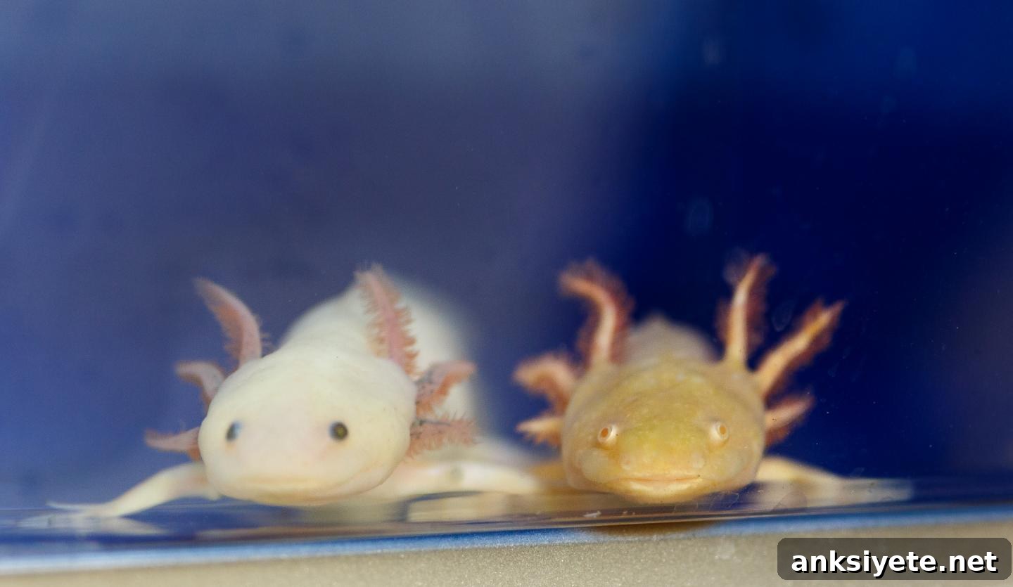
Summary: Researchers have identified gene partnerships in the axolotl that enable the neural tube and nerve fibers to regenerate after severe spinal cord injury. Those same genes exist in humans but are activated differently, which helps explain why humans form scar tissue instead of regenerating.
Source: Marine Biological Laboratory
Researchers are closing in on why some vertebrates regenerate their spinal cords while others, including humans, form scars and suffer permanent loss of function.
Scientists at the Marine Biological Laboratory (MBL) have discovered specific gene interactions in the axolotl salamander that drive a pro-regenerative response in glial cells after spinal cord injury. The team found that these interactions allow the axolotl to rebuild its neural tube and reconnect nerve fibers so the animal regains both motor and sensory function. Crucially, the same genes exist in humans but are engaged differently following injury, which appears to influence whether an injured spinal cord regenerates or becomes scarred. The study appears in Nature Communications Biology.
Axolotls are well known for their exceptional regenerative capacity. When the animals sustain a spinal cord lesion, certain glial cells proliferate and migrate to rebuild missing tissue and create an environment that supports axon regrowth. Karen Echeverri, an associate scientist in the Eugene Bell Center for Regenerative Biology and Tissue Engineering at MBL, explains that the goal of the research was to determine what molecular differences steer axolotls toward regeneration rather than scar formation.
Previous work by Echeverri and colleagues showed that the gene c-Fos is upregulated in glial cells of both axolotls and humans after spinal cord injury. However, c-Fos does not act alone. As an obligate heterodimer, c-Fos must pair with a partner protein to regulate downstream gene expression. The identity of that partner turns out to be a key determinant of the injury response.
In humans, the injury response typically pairs c-Fos with the partner c-Jun, a combination associated with reactive gliosis and scar formation. In axolotls, the researchers found that c-Fos pairs predominantly with JunB after injury. That different pairing correlates with a pro-regenerative program in axolotl glial cells rather than the reactive, scarring response seen in mammalian tissue.
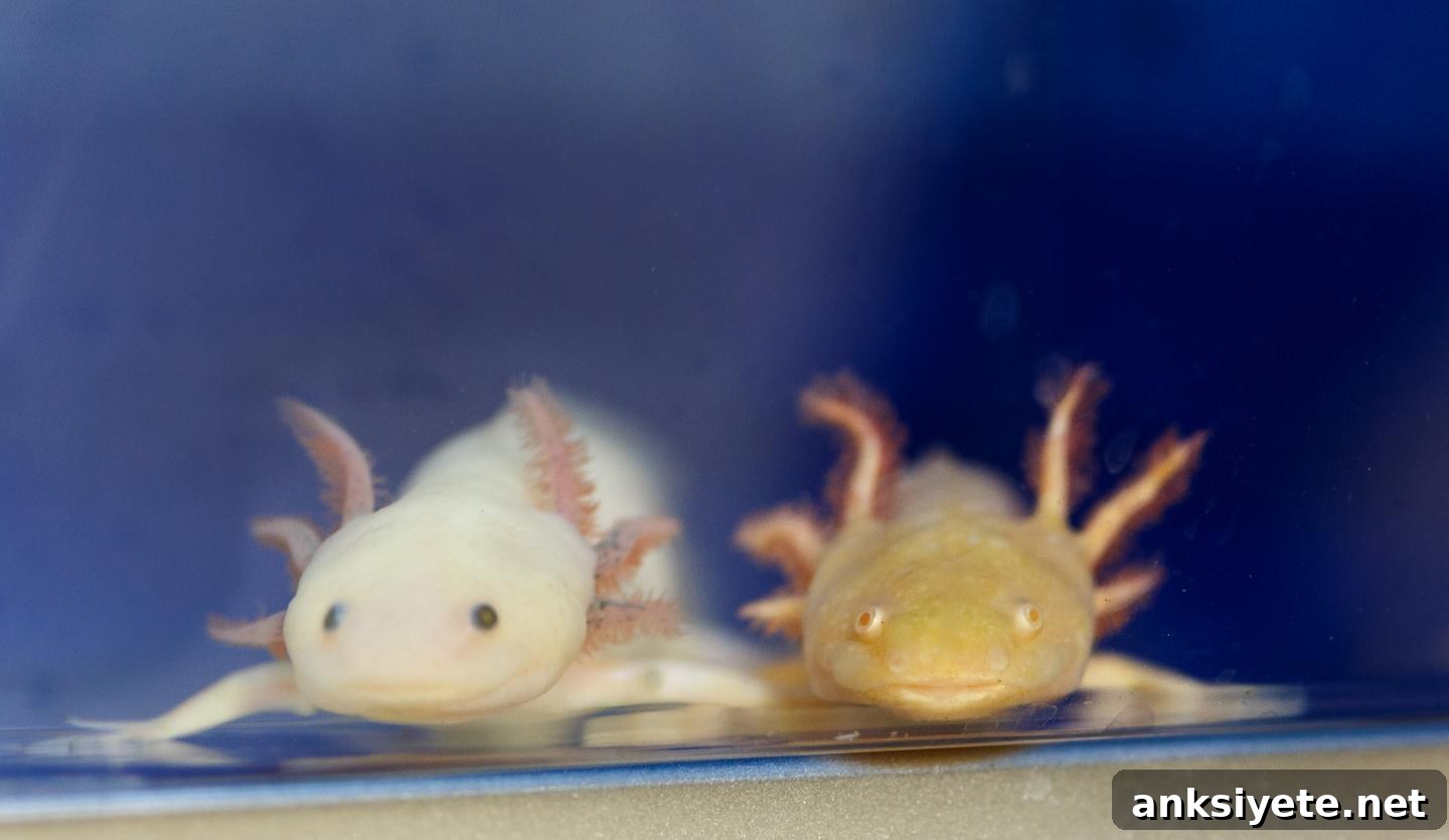
The researchers traced this difference in pairing to microRNAs, small regulatory RNAs that modulate gene expression. In axolotls, injury triggers upregulation of miR-200a in glial cells. miR-200a suppresses c-Jun expression, limiting the availability of that partner and biasing c-Fos to partner with JunB instead. When the team experimentally altered microRNA regulation to force the c-Fos/c-Jun pairing in axolotls, the salamanders no longer regenerated a functioning spinal cord and instead developed scar tissue similar to the human response.
These experiments demonstrate that the genes controlling regeneration are conserved between axolotls and humans; the critical difference is how those genes are regulated immediately after injury. “It’s all about who you partner with directly after injury, and how that drives you toward either regeneration or forming scar tissue,” Echeverri summarizes. The finding highlights gene partnership and microRNA control as potential molecular switches that tip the balance between productive regeneration and damaging scarring.
Understanding the axolotl’s pro-regenerative program — and the regulatory mechanisms that prevent the same outcome in mammals — has clear translational relevance. If researchers can determine whether altering microRNA or AP-1 component interactions produces a comparable pro-regenerative response in human cells, those pathways could become targets for therapies to improve recovery after spinal cord injury. Such advances might also inform treatments for certain neurodegenerative conditions where promoting regeneration or reducing reactive gliosis would be beneficial.
MBL has a long history of basic research in nontraditional model organisms leading to insights relevant to human health. Studies of animals with extraordinary biological traits often reveal conserved molecular mechanisms that can be manipulated or mimicked in other species. This work on axolotl spinal cord regeneration is an example of how fundamental discovery research can point to new strategies for addressing human medical challenges.
Written by Stephanie McPherson
Source: Marine Biological Laboratory
Media Contact: Diana Kenney – Marine Biological Laboratory
Publisher: Neuroscience News, reporting on MBL research
Image credit: Dee Sullivan
Original research: AP-1cFos/JunB/miR-200a regulate the pro-regenerative glial cell response during axolotl spinal cord regeneration. DOI: 10.1038/s42003-019-0335-4
Funding: National Institutes of Health
Abstract (summary)
Salamanders can functionally regenerate after spinal cord transection. In axolotl spinal cord injury, GFAP+ glial cells proliferate and migrate to replace the missing neural tube and create an environment permissive for axon regeneration. The molecular pathways that regulate this pro-regenerative glial response were previously unclear. The study shows axolotl glial cells upregulate an AP-1 complex composed of c-Fos and JunB after injury, promoting regeneration. Injury-induced upregulation of miR-200a suppresses c-Jun expression, preventing formation of the c-Fos/c-Jun complex associated with reactive gliosis. Inhibiting miR-200a impairs axon regrowth and alters expression of genes linked to gliosis, extracellular matrix remodeling, and axon guidance. These findings identify miR-200a as a key inhibitor of reactive gliosis during axolotl spinal cord regeneration.