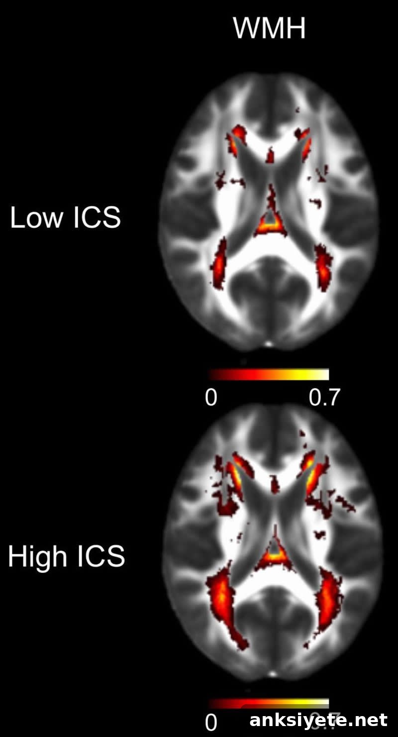
Summary: Six inflammatory proteins measured in blood can help assess an individual’s risk of cerebral small vessel disease (CSVD), a form of brain small-vessel injury linked to higher risk of stroke and dementia, including Alzheimer’s disease.
Source: UCLA
UCLA-led research shows that blood levels of six proteins can indicate a person’s risk for cerebral small vessel disease (CSVD), a common cause of white matter damage that affects an estimated 11 million older adults in the U.S. CSVD contributes to cognitive decline and stroke but currently is most reliably identified by brain MRI.
“Our goal is to develop a new, quantitative blood test that clinicians can use to evaluate brain vascular health and identify people at risk for cerebral small vessel disease,” said Dr. Jason Hinman, a UCLA assistant professor of neurology and the study’s lead author. The findings appear in the journal PLOS ONE.
CSVD primarily damages the brain’s white matter—the myelin-rich regions that allow rapid communication between neurons. Over time, the tiny blood vessels that supply white matter may become compromised. As myelin breaks down, signal transmission slows, which can cause walking difficulties, memory problems, and other cognitive impairments. Complete blockage of these small vessels can also trigger ischemic stroke.
The condition is associated with an increased risk of multiple types of dementia, including Alzheimer’s disease. Clinically, CSVD is usually detected on MRI after symptoms appear or after a stroke, and it accounts for roughly one-quarter of strokes in the United States. Because early symptoms are often subtle—such as mild gait disturbance or memory lapses—many cases remain undiagnosed and are mistaken for normal aging.
In this study, Hinman and colleagues tested a hypothesis that inflammatory proteins implicated in CSVD would be measurable in blood. They focused on six immune-related proteins, anchored around the cytokine interleukin-18 (IL-18), reasoning that an IL-18-centered inflammatory network might reflect cerebrovascular white matter injury.
The team measured these six proteins in blood samples from 167 volunteers (mean age 76.4) with either normal cognition or mild cognitive impairment. Of these participants, 110 underwent standard MRI and 49 received diffusion tensor imaging (DTI), a more sensitive scan for early white matter changes.
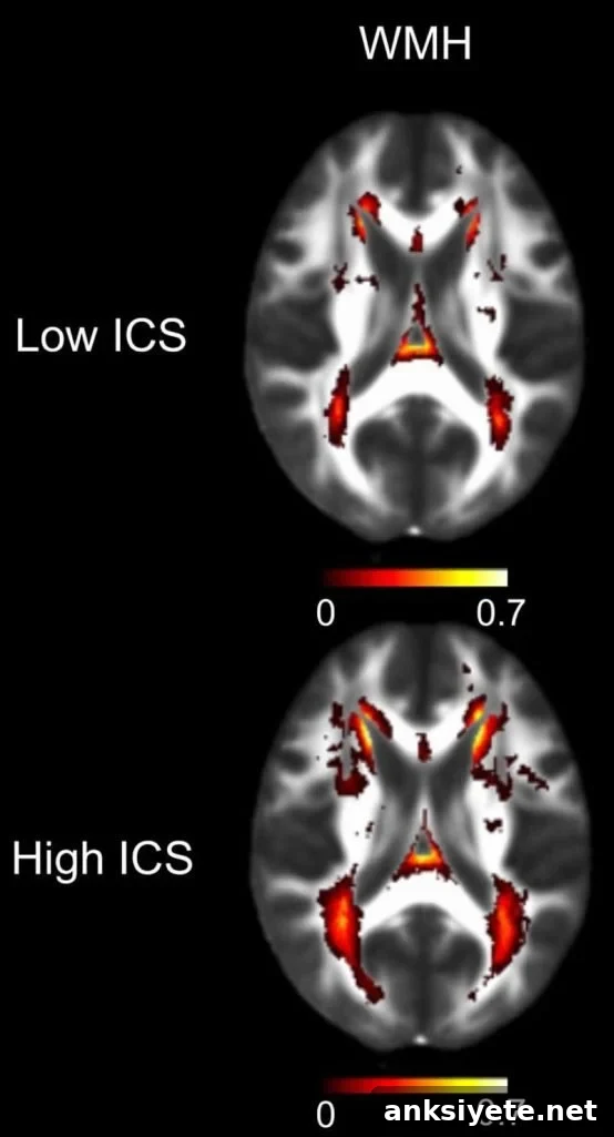
The researchers found that participants with MRI or DTI evidence of CSVD had significantly higher levels of the six inflammatory proteins. Individuals with above-average levels were roughly twice as likely to show CSVD on MRI and about 10% more likely to display very early white matter abnormalities on DTI. Furthermore, each additional vascular risk factor—such as hypertension, diabetes, or a prior stroke—was associated with approximately a twofold increase in the inflammatory composite score on average.
To validate these observations in a separate, higher-risk group, the team applied the blood test to 131 people who presented to a UCLA Health emergency department with symptoms concerning for stroke. In this cohort, the inflammatory protein measurements again correlated with white matter changes identified on MRI.
“I was pleasantly surprised that we were able to associate bloodstream inflammation with CSVD in two fairly different populations,” Hinman said.
Currently, MRI reports typically categorize white matter changes in broad terms—mild, moderate, or severe. The blood-based inflammatory composite offers a more quantitative measure of CSVD burden, which could help clinicians monitor disease progression and identify candidates for prevention strategies or clinical trials of therapies targeting vascular brain injury.
“We hope this work encourages more quantitative approaches to CSVD assessment, so clinicians and researchers can better target interventions,” Hinman added.
The blood test is not yet commercially available.
The study’s first author is Marie Altendahl, a UCLA medical student who conducted the work while at UC San Francisco. Other UCLA-affiliated contributors include former undergraduate researcher Visesha Kakarla and former postdoctoral fellow Guanxi Xiao. Additional authors are affiliated with UC San Francisco and UC Davis.
Funding: This research was supported by the National Institutes of Health, the American Heart Association, the MarkVCID consortium, and the Lillian R. Gleitsman Foundation.
Source:
UCLA
Media Contacts:
Marrecca Fiore – UCLA
Image Source:
Image credit: UCLA Health.
Original Research: Open access — “An IL-18-centered inflammatory network as a biomarker for cerebral white matter injury”. Marie Altendahl, Pauline Maillard, Danielle Harvey, Devyn Cotter, Samantha Walters, Amy Wolf, Baljeet Singh, Visesha Kakarla, Ida Azizkhanian, Sunil A. Sheth, Guanxi Xiao, Emily Fox, Michelle You, Mei Leng, David Elashoff, Joel H. Kramer, Charlie Decarli, Fanny Elahi, Jason D. Hinman. PLOS ONE. DOI: 10.1371/journal.pone.0227835.
Abstract
An IL-18-centered inflammatory network as a biomarker for cerebral white matter injury
Chronic systemic sterile inflammation is implicated in the development of cerebrovascular disease and white matter injury, but noninvasive blood markers for risk stratification and in vivo assessment are limited. This study evaluated whether an interconnected set of inflammatory biomarkers centered on IL-18—previously linked to white matter lesions—could detect established and early white matter changes in two at-risk populations. In a cohort of 167 older adults (mean age 76) who completed cognitive testing, physical exams, blood collection, and MR imaging including DTI, the researchers measured white matter hyperintensities (WMH) and free water (FW). Serum levels of six inflammatory molecules (MPO, GDF-15, RAGE, ST2, IL-18, and MCP-1) were quantified and combined into a log-transformed, population mean-adjusted inflammatory composite score (ICS). Adjusted models that accounted for age and total intracranial volume showed that ICS was significantly associated with WMH (β = 0.222, p = 0.013), FW (β = 0.3, p = 0.01), and with the number of vascular risk factor diagnoses (r = 0.36, p < 0.001). In a second cohort of 131 patients evaluated for suspected stroke, an unbiased principal component analysis of 11 inflammatory biomarkers identified a factor strongly correlated with the six-component ICS and associated with WMH after adjusting for age and gender (p = 0.027). These results indicate that an IL-18-driven network of inflammatory molecules is linked to both overt and antecedent white matter injury from cerebrovascular disease and may serve as a promising peripheral biomarker for vascular white matter damage.