
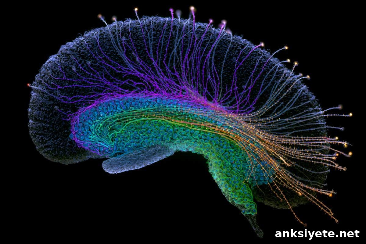
Summary: A new single-cell multiomic study finds that the human brain undergoes coordinated shifts in gene regulation and three-dimensional genome architecture beginning in midlife. By profiling individual cells across the adult lifespan in the hippocampus—a region essential for learning and memory—researchers mapped structural genome breakdown and system-level remodeling that help explain why aging is the primary risk factor for neurodegenerative disease.
Key Facts
- Midlife Microglial Replacement: Between about ages 50 and 75, embryonically derived, resident microglia decline and are largely replaced by cells with molecular signatures resembling peripheral blood immune cells, which display elevated pro-inflammatory profiles.
- 3D Genome Architecture Erosion: Across multiple hippocampal cell types, investigators observed progressive weakening of three-dimensional genome organization, identifying structural genomic decay as a hallmark of brain aging.
- Vascular and Barrier Compromise: Populations that help maintain blood-brain barrier integrity decline with age, increasing vulnerability to circulating toxins and neuroinflammation.
- Coordinated Multisystem Remodeling: Aging in the human hippocampus involves dynamic, coordinated changes among neuronal, vascular, and innate immune networks rather than simple passive decline.
- Part of NIH 4D Nucleome Initiative: This study is one of six papers from the NIH 4D Nucleome Consortium published together to map the genome’s spatial and temporal organization in human health and disease.
Source: NYGC
Overview: Using advanced single-cell and multiomic methods, researchers examined gene regulation, chromatin state, DNA methylation, and three-dimensional genome architecture in individual hippocampal cells collected across the adult lifespan. The analysis produced one of the most comprehensive profiles to date of how genome regulation changes during human brain aging.
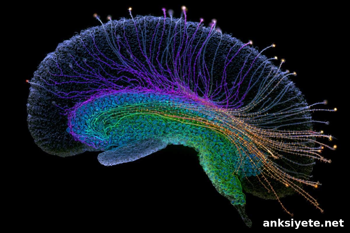
A central finding is a striking shift in the brain’s immune landscape during midlife: embryonic microglia decline substantially and are replaced by a cell population that carries epigenetic and transcriptional signatures similar to monocytes from the blood. These replacement cells show heightened inflammatory programs, implicating them as contributors to chronic neuroinflammation in aging.
Concurrently, the study documents a marked reduction in astrocyte subtypes that support synaptic function and maintain the blood-brain barrier. Those astrocytes also show signs of metabolic stress and reduced mitochondrial gene expression, suggesting energy dysfunction may underlie their loss and further compromise brain homeostasis.
On the level of genome organization, investigators detected a global weakening of higher-order chromatin structure—loss of integrity in topologically associating domains and increased aberrant interchromosomal contacts—across diverse cell types. These structural changes align with epigenetic alterations and shifts in gene expression, revealing widespread rewiring of regulatory programs with age.
“Microglia are essential for brain homeostasis,” said Bing Ren, PhD, a corresponding author. “When microglial functions erode and are replaced by pro-inflammatory cells, toxic debris can accumulate and trigger persistent inflammatory responses that may accelerate neurodegenerative processes.”
Nathan Zemke, Director of Single-cell Genomics at UC San Diego’s Center for Epigenomics, noted that integrating gene expression with DNA methylation and 3D genome mapping was critical to detect lineage changes that transcriptional data alone would miss. The DNA methylation signatures preserved lineage history, revealing the monocyte-like origin of replacement microglia.
Xiangmin Xu, PhD, emphasized that aging should be viewed as an active, coordinated remodeling process: “These results show aging reshapes immune, vascular, and neuronal systems in ways that create new vulnerabilities. Understanding these coordinated changes opens the door to targeted interventions to preserve circuits and cognition.”
Key Questions Answered:
A: Between roughly ages 50 and 75, embryonically derived resident microglia decline and are replaced by microglia-like cells with blood-derived immune signatures. These cells carry stronger pro-inflammatory gene programs and likely contribute to chronic neuroinflammation.
A: 3D genome architecture describes how DNA folds and organizes spatially in the nucleus to regulate gene activity. Its deterioration disrupts normal gene regulation, contributes to cellular dysfunction, and undermines the maintenance of brain tissue.
A: The work reframes aging as a dynamic, coordinated process that reshapes immune, vascular, and neuronal systems starting in midlife. Targeting specific epigenomic and structural changes could enable earlier interventions to preserve brain function and lower neurodegenerative risk.
Editorial Notes:
- Edited by a Neuroscience News editor.
- Original journal paper was reviewed in full.
- Additional context was added by editorial staff.
About this research
Author: Jessica Leitner
Source: New York Genome Center
Contact: Jessica Leitner – New York Genome Center
Image credit: Neuroscience News
Original Research: Open access. “New Study Revises Understanding of Brain Immune Cells During Human Aging” published in Science. DOI provided in the original paper.
Abstract
New Study Revises Understanding of Brain Immune Cells During Human Aging
INTRODUCTION
Aging is the strongest risk factor for neurodegenerative disease and is accompanied by memory loss and chronic brain inflammation. The hippocampus is especially vulnerable. Previous work identified age-related changes in gene expression—more inflammatory signaling and reduced synaptic function—but expression data alone do not capture underlying epigenetic and structural drivers of dysfunction.
RATIONALE
To define molecular mechanisms of hippocampal aging, the study integrated single-cell measurements of gene expression, chromatin accessibility, DNA methylation, and 3D genome organization across the adult lifespan. This multiomic approach reveals coordinated, cell type–specific epigenetic and architectural changes that single-modality studies can miss.
RESULTS
Aging produced coordinated and often nonlinear regulatory changes across cell types, with a pronounced midlife transition. Embryonic, brain-resident microglia were progressively replaced by cells with DNA methylation and chromatin features resembling circulating monocytes; these monocyte-like microglia exhibit proinflammatory transcriptional and 3D genome signatures. Astrocyte populations that support synapses and the blood-brain barrier declined, accompanied by reduced expression of mitochondrial and metabolic genes and elevated stress signatures. Genome architecture weakened broadly, with loss of domain integrity and increased aberrant interchromosomal interactions, coinciding with epigenetic shifts and transcriptional rewiring.
CONCLUSION
Aging reprograms both the cellular composition and regulatory architecture of the human hippocampus. Replacement of embryonic microglia by a proinflammatory, monocyte-like population, loss of supportive astrocytes, and widespread alterations in 3D genome structure emerge as key hallmarks of brain aging. Together, these coordinated changes provide a mechanistic framework for age-related cognitive decline and heightened vulnerability to neurodegenerative disease.