
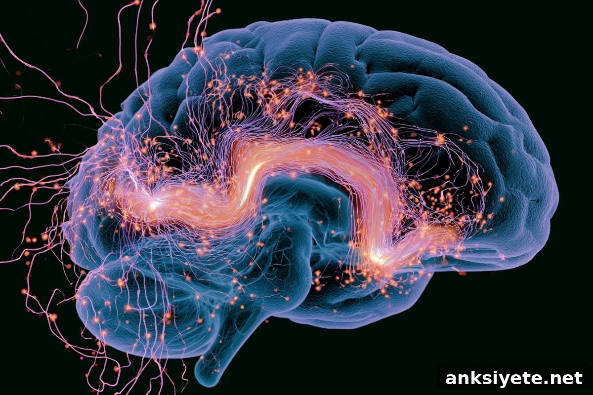
Summary: Scientists have uncovered a subcortical mechanism that explains why memory accessibility can change from moment to moment. Using real-time neural monitoring, optogenetics, and deep-brain calcium imaging in mice, researchers found that slow, spontaneous fluctuations in hypothalamic histamine neurons act as a gatekeeper for retrieval. High baseline histaminergic activity just before a cue primes downstream memory circuits—particularly within the basolateral amygdala—so a stored memory is readily accessed. When histamine activity is low, the identical cue often fails to evoke the memory pattern even though the memory trace itself remains intact.
This discovery reframes retrieval failures: they are frequently due to transient internal brain states that make memories temporarily inaccessible, not to permanent loss of the memory trace. The findings identify a specific, manipulable subcortical process that sets the brain’s readiness to recall information.
Key Facts
- Memory trace versus accessibility: Rather than erasure of engrams, retrieval variability often reflects continuous internal-state fluctuations that determine whether an intact memory can be read out at any given moment.
- Histamine neurons as a timing signal: Histamine-producing neurons in the tuberomammillary nucleus (TMN) of the hypothalamus—known for regulating wakefulness—show slow spontaneous waves of activity that rise and fall over tens of seconds and project widely to memory-related regions, including cortex, hippocampus, and amygdala.
- 40% gating effect: In closed-loop experiments that presented a learned auditory cue precisely at the peak or trough of histaminergic activity, mice were about 40% more likely to produce memory-guided behavior when cues occurred during high-histamine states.
- Optogenetic causal evidence: Direct optogenetic suppression of histamine neuron firing immediately before a memory cue blocked retrieval behavior; optogenetic activation restored successful recall—demonstrating causality, not just correlation.
- Specific priming distinct from general arousal: Manipulating histamine neurons did not change locomotion, basic auditory thresholds, pupil size, or innate reward consumption, indicating a specific priming role for memory circuits rather than a broad arousal effect.
- Amygdala network stabilization: Deep-brain calcium imaging in the basolateral amygdala (BLA) showed that high histaminergic states stabilized the precise population firing pattern associated with the learned cue; suppression of histamine activity weakened and destabilized that pattern.
- Clinical implications: Mapping this histaminergic priming axis suggests new diagnostic and therapeutic approaches to stabilize moment-to-moment memory access in aging and neurodegenerative conditions that feature fluctuating cognition, such as Alzheimer’s disease and related dementias.
Source: Nagoya City University
The same memory can feel vivid and accessible one moment and stubbornly out of reach the next—even though the underlying trace is preserved. A research group led by Professor Hiroshi Nomura at the Institute of Brain Science, Nagoya City University Graduate School of Medical Sciences, identified a neural mechanism that may explain these rapid changes in accessibility.
The team demonstrated that infraslow fluctuations in hypothalamic histamine neurons influence whether a memory will be expressed when a cue appears. When histamine neuron activity rose before a cue, mice were more likely to show the learned response; when activity dipped, identical cues often failed to elicit the behavior.
“Our results show that a failure to recall does not always mean the memory is gone,” said Hiroshi Nomura, senior author. “Instead, the brain can enter brief states in which stored memories are difficult to access.”
Histamine neurons in the TMN are best known for promoting wakefulness and influencing peripheral systems like allergy responses. They also send wide-ranging projections to memory hubs. Recording from awake mice, the research team observed slow, infraslow dynamics (about 0.05–0.1 Hz) in histamine neuron firing that tracked a combined brain–body state reflected in cortical activity, pupil size, and facial movements.
In behavioral tests, mice learned to associate a sound with a sugar-water reward and responded by licking when the sound played. Trials with higher pre-cue histamine activity were much more likely to produce significant cue-evoked licking, suggesting that histamine activity primes downstream circuits in advance of the cue.
Using a closed-loop system that presented the cue during naturally high or low histamine states, the researchers found roughly a 40% boost in memory-guided responses during high states. Optogenetic manipulations confirmed causality: brief activation of histamine neurons increased recall, while brief suppression reduced it. Crucially, these changes occurred without altering basic sensory thresholds, motor ability, or immediate reward responses, indicating a targeted effect on memory accessibility.
Calcium imaging in the basolateral amygdala revealed the downstream mechanism: when mice successfully expressed the learned memory, BLA networks reliably reproduced the exact population pattern established during learning. When histamine activity was suppressed before the cue, that population pattern degraded and became unreliable, blocking effective recall.
Overall, the data support a “priming-state” model in which infraslow histaminergic dynamics set a preparatory brain state. That state makes it more or less likely that an incoming cue will trigger the precise neural pattern required for recall.
Nomura emphasized the conceptual shift: “Rather than treating recall as a simple readout of a stored trace, we must consider internal brain state as a gate that determines whether the trace becomes accessible at a given moment.”
Because this study used a reward-based memory task in mice, further work will test whether histaminergic priming shapes other memory types—such as fear, spatial, or social memory—and whether similar fluctuations contribute to everyday memory variability in humans. The research also suggests a promising target for therapies aimed at stabilizing memory access in conditions marked by fluctuating cognition, including aging and dementia.
Key Questions Answered:
A: No. This study shows that a memory can remain intact but be temporarily inaccessible because of internal brain-state fluctuations. Slow changes in histaminergic activity act like a dimmer switch that governs whether a memory trace is open for readout at a given moment.
A: Histamine acts as a subcortical priming signal. When histamine neuron firing is high just before a cue, it stabilizes downstream circuits—such as the basolateral amygdala—so the incoming cue can reliably recreate the neural pattern needed for recall.
A: By identifying a biological target—the histaminergic priming axis—researchers can explore treatments to stabilize these infraslow waves and keep the brain in a more consistent “ready-to-recall” state, potentially reducing unpredictable fluctuations in everyday memory for patients with Alzheimer’s disease and related dementias.
Editorial Notes:
- This article was edited by a Neuroscience News editor.
- Journal paper reviewed in full.
- Additional context added by staff.
About this memory and neuroscience research news
Author: Hirano Anna
Source: Nagoya City University
Contact: Hirano Anna – Nagoya City University
Image: Image credited to Neuroscience News
Original Research: Open access. “Infraslow histaminergic dynamics govern priming states to gate moment-to-moment memory accessibility” by Yoshikazu Morishita, Yuki Takamura, Kyoka Nishimura, Natsuko Hitora-Imamura, Masabumi Minami, and Hiroshi Nomura. Neuron. DOI: 10.1016/j.neuron.2026.05.019
Abstract
Infraslow histaminergic dynamics govern priming states to gate moment-to-moment memory accessibility
Memory expression can vary in response to identical cues, implying that ongoing brain states bias accessibility. The cellular and circuit mechanisms behind these state-dependent fluctuations have been unclear. This study shows that spontaneous pre-cue activity of histaminergic neurons in the hypothalamic tuberomammillary nucleus modulates reward-associative memory expression in mice. TMN histaminergic activity displayed infraslow dynamics (0.05–0.1 Hz) that tracked an integrated brain–body state.
Closed-loop cue presentation during high histaminergic states enhanced memory expression. Brief optogenetic activation or inhibition of these neurons before the cue bidirectionally altered memory expression, and activation of histaminergic terminals in the basolateral amygdala was sufficient to improve recall. Histaminergic inhibition before the cue impaired the BLA’s cue-evoked population response. Thus, ongoing histaminergic activity exerts an infraslow, state-setting influence that primes BLA circuits for robust cue responses and modulates moment-to-moment memory accessibility.