
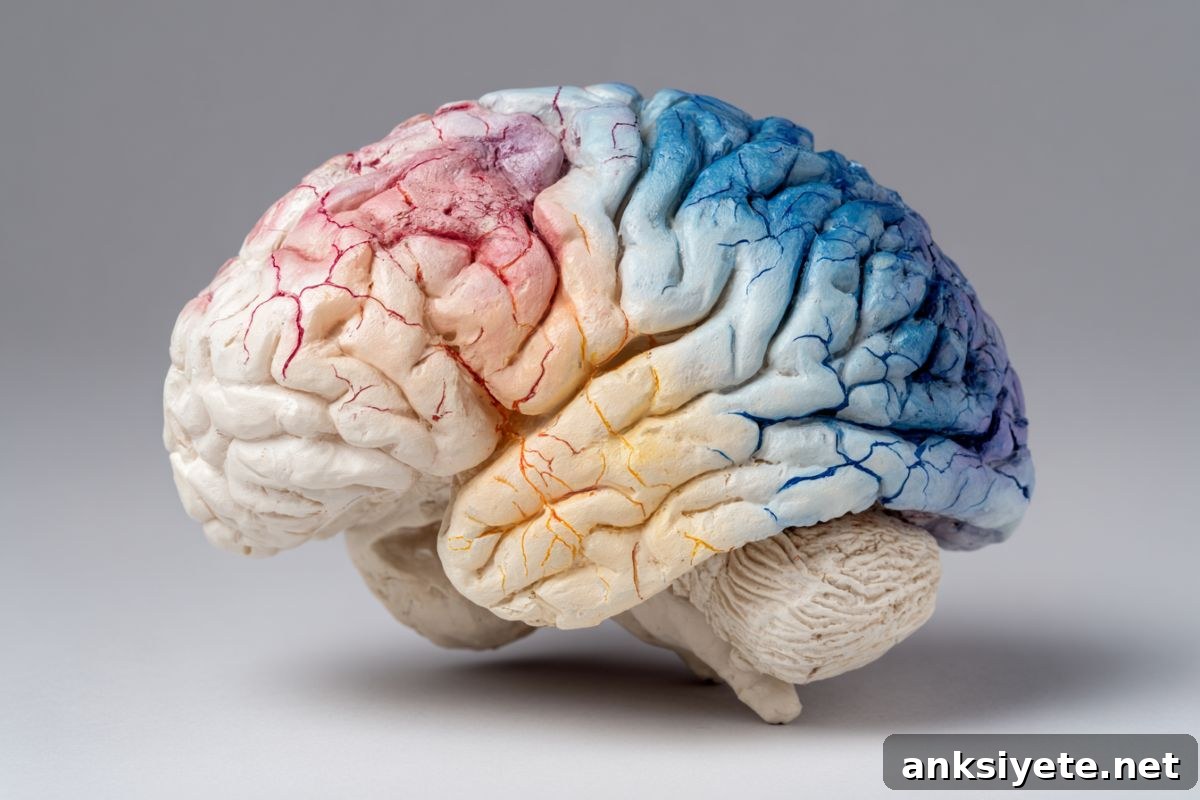
Summary: In a large international study, researchers identified an unexpected pattern of neuroplasticity after stroke. By applying deep learning to MRI scans from over 500 stroke survivors across eight countries, they observed that while the damaged hemisphere shows accelerated aging, the undamaged hemisphere often develops structural features that appear “younger.”
This regional rejuvenation is most pronounced in areas involved in motor planning and attention, suggesting the healthy side of the brain reorganizes to compensate for loss of function.
Key Facts
- Brain-PAD marker: The team used AI-derived Brain-Predicted Age Difference (brain-PAD) to estimate biological age from MRI scans. A lower (younger) brain-PAD in undamaged regions served as an indicator of structural reorganization.
- The contralesional shift: Stroke survivors with the most severe movement impairments showed the most pronounced “youthful” structural patterns in the hemisphere opposite the lesion, especially in the frontoparietal network.
- Global collaboration: The study was coordinated by the ENIGMA Stroke Recovery Working Group, harmonizing data from 34 sites to assemble the largest stroke neuroimaging dataset to date.
- Paradoxical adaptation: The apparent rejuvenation does not necessarily imply full motor recovery. Instead, it reflects compensatory remodeling of healthy brain tissue to support lost motor functions.
Source: USC
Overview: A new study published in The Lancet Digital Health by scientists at the USC Mark and Mary Stevens Neuroimaging and Informatics Institute (Stevens INI) reports that, after severe stroke, undamaged regions of the brain can show structural signatures consistent with a “younger” brain. These changes appear to reflect adaptive reorganization aimed at supporting impaired motor systems.
As part of the ENIGMA Stroke Recovery Working Group, researchers analyzed structural MRI scans from more than 500 individuals with chronic unilateral stroke collected across 34 cohorts in eight countries. They used deep learning models trained on large population datasets to estimate regional brain age in each hemisphere.
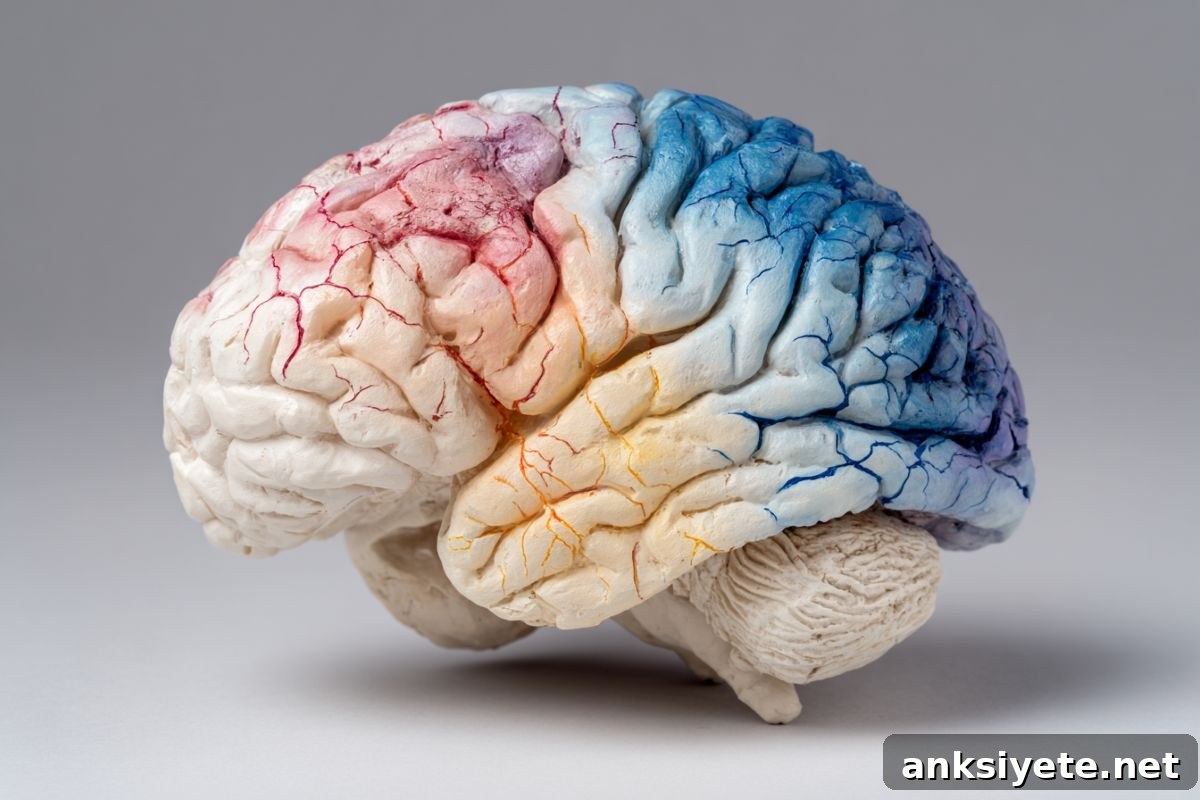
Using a graph convolutional network trained on tens of thousands of MRI scans, the investigators estimated the biological age of 18 functional brain subregions. The difference between predicted brain age and chronological age (brain-PAD) served as a sensitive biomarker of regional structural health.
Lead authors describe a clear pattern: larger lesions are associated with older-appearing tissue in the damaged (ipsilesional) hemisphere, while more severe motor deficits are associated with younger-appearing tissue in specific contralesional regions, most notably the frontoparietal network—a system critical for motor planning, attention, and coordination.
“When the damaged motor system cannot perform its role, the opposite hemisphere appears to reorganize and take on greater responsibility,” said Hosung Kim, PhD, co-senior author and associate professor of research neurology at the Keck School of Medicine of USC. “This shows a form of structural neuroplasticity that conventional imaging often misses.”
The study linked regional brain-PAD values to clinical motor scores. Participants with persistent, severe motor impairments—those still limited in movement after more than six months of rehabilitation—showed the most pronounced contralesional “youthful” signatures. This suggests an “emergency” adaptation: healthy networks strengthen structural connectivity in response to severe damage.
By harmonizing MRI and clinical data across dozens of cohorts, the ENIGMA team created a dataset large enough to detect subtle, region-specific effects. Arthur W. Toga, PhD, director of the Stevens INI, noted that pooling international data and applying advanced AI made these discoveries possible and could guide more targeted rehabilitation approaches in the future.
Planned follow-up work will track patients longitudinally from the acute to chronic phases to see how regional brain aging evolves over time. Understanding the timeline and location of these plastic changes could enable clinicians to tailor therapies that reinforce beneficial reorganization and improve long-term outcomes.
Key Questions Answered:
A: “Younger” in this context refers to structural features—greater cortical thickness, denser connectivity, or patterns of regional organization—detected by AI models. These changes reflect increased recruitment and reinforcement of healthy networks, resembling the structural profile typically seen in younger brains.
A: Not necessarily. The most pronounced “younger” patterns appeared in patients with the most severe impairments, suggesting this adaptation is a response to substantial loss rather than a sign of rapid or full recovery.
A: Brain-PAD and regional age maps could inform personalized rehabilitation by identifying which contralesional networks are attempting to compensate, allowing therapists to target and strengthen those pathways with tailored interventions.
Editorial Notes:
- This article was edited by a Neuroscience News editor.
- The journal paper was reviewed in full by editorial staff.
- Additional context and clarification were added by our team.
About this neurology and stroke research news
Author: Laura LeBlanc
Source: USC
Contact: Laura LeBlanc – USC
Image: Image credit to Neuroscience News
Original Research: Closed access. “Associations between contralesional neuroplasticity and motor impairment through deep learning-derived MRI regional brain age in chronic stroke (ENIGMA): a multicohort, retrospective, observational study” by Gilsoon Park et al., Lancet Digital Health. DOI: 10.1016/j.landig.2025.100942
Abstract
Associations between contralesional neuroplasticity and motor impairment through deep learning-derived MRI regional brain age in chronic stroke (ENIGMA): a multicohort, retrospective, observational study
Background
Stroke produces lasting structural and functional changes that influence motor recovery. Brain-predicted age difference (brain-PAD) has emerged as a sensitive biomarker for post-stroke sensorimotor and cognitive status. Prior work linked higher global brain-PAD with worse motor outcomes, but relationships among focal lesion burden, regional brain age, and motor impairment remained unclear. This study examined how lesion load and regional brain-PAD in both hemispheres associate with motor outcomes in chronic unilateral stroke and sought key predictors of motor impairment.
Methods
This multicohort, retrospective study included individuals more than 180 days post unilateral stroke from the ENIGMA Stroke Recovery dataset. The team trained a regional brain age prediction model using UK Biobank participants. Structural T1-weighted MRI scans produced regional brain-PAD estimates for 18 predefined functional subregions using a graph convolutional network. Lesion load per region was computed from lesion overlap. Linear mixed-effects models tested associations among lesion size, local lesion load, and regional brain-PAD. Machine learning classifiers evaluated motor outcome prediction using lesion loads and regional brain-PADs, and structural equation modeling explored directional relationships among corticospinal tract lesion load, ipsilesional brain-PAD, motor outcomes, and contralesional brain-PAD.
Findings
The study analyzed 501 individuals from 34 cohorts and 17,791 UK Biobank participants for training. Larger total lesion size correlated with older-appearing tissue in the ipsilesional hemisphere across multiple regions and with younger-appearing tissue in contralesional ventral attention and language regions. Similar patterns emerged for local lesion loads, with the salience network lesion burden influencing regional brain-PAD bilaterally. Machine learning identified corticospinal tract lesion load, salience network lesion load, and contralesional frontoparietal regional brain-PAD as top predictors of motor outcomes. Structural equation modeling suggested higher corticospinal tract lesion load leads to poorer motor outcomes, which are associated with younger contralesional brain age—consistent with compensatory contralesional remodeling in severe impairment.
Interpretation
These results indicate larger stroke lesions accelerate aging in the damaged hemisphere while paradoxically slowing apparent aging in contralesional regions, implying compensatory neuroplasticity. Regional brain age measures may serve as biomarkers of adaptive reorganization and help guide targeted interventions to improve motor recovery after stroke.
Funding
US National Institutes of Health.