
Summary: Researchers used neuroimaging to identify reliable brain patterns linked to two distinct empathic responses—compassionate care and empathic distress—that are consistent and predictable across people.
Source: Cell Press.
When we see others suffer, we can respond in different ways: we may feel distressed as we imagine another’s pain, or we may experience empathic care—compassion and a desire to help. A study published in Neuron on June 8 used naturalistic stories of human hardship and functional magnetic resonance imaging (fMRI) to show that these two forms of empathy correspond to distinct, distributed patterns of brain activity that are consistent across individuals.
“Feelings of empathy are virtues we want to cultivate personally and socially,” says first author Yoni Ashar, a graduate student in Tor D. Wager’s lab at the University of Colorado, Boulder. “Understanding how these emotions arise in the brain could help us foster more compassion while reducing harmful distress.”
Ashar offers a simple example from everyday life: when his toddlers cry, he sometimes mirrors their upset and experiences empathic distress. At other moments he intentionally shifts to empathic care—calm concern and support—rather than becoming overwhelmed.
To investigate how these emotions map onto brain activity, the research team recruited 66 adults who listened to 24 short, true stories about people facing hardship while inside an fMRI scanner. The stories were chosen to evoke natural emotional responses—for instance, a young person overcoming addiction and later helping others recover. Unlike prior studies that used static images, this naturalistic approach mirrors how we typically encounter others’ suffering in real life.
Participants later listened to the same narratives outside the scanner and provided continuous ratings of their moment-by-moment feelings of distress and care as each story unfolded. The researchers used those self-reports to train and validate brain-based markers that predict the intensity of empathic care and empathic distress from fMRI activity patterns.
Rather than localizing empathy to a single brain region, the study found that empathic feelings are represented by widely distributed neural patterns. “The brain is not a simple modular system where a single region manages empathy,” says Tor Wager. “Empathic states recruit multiple interacting systems across the brain.”
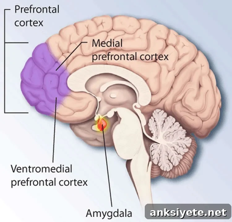
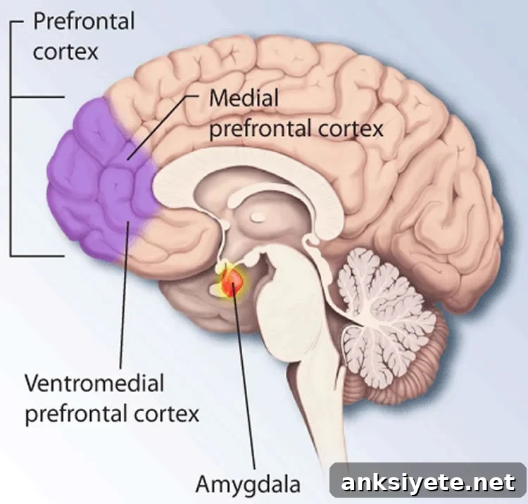
The neural signature of empathic care overlapped with brain areas normally associated with value and reward processing, including the ventromedial prefrontal cortex (vmPFC) and medial orbitofrontal cortex (mOFC). In contrast, empathic distress corresponded to activity in regions linked to mirroring and somatic simulation—such as the premotor cortex and primary and secondary somatosensory cortices—areas that help people internally simulate another person’s sensations and actions.
These neural patterns were remarkably consistent across participants. The researchers were able to predict, from brain activity alone, empathic responses in individuals who had not been included in the model training. “There is individual variation in when and how people experience care or distress, but the neural systems activated during those states are similar across people,” Ashar explains.
To further characterize the emotional content of these empathic states, a separate sample of 200 adults provided continuous ratings of basic emotions—sadness, disgust, anger, fear, negativity, positivity, and happiness—while listening to the same stories. Mapping empathic markers onto these basic feelings revealed that empathic care reflects a mixed-valence state combining warmth and elements of sadness and positivity, whereas empathic distress aligns primarily with negative emotions such as sadness, anger, fear, and disgust.
Beyond mapping emotions, the fMRI markers had behavioral relevance: brain patterns associated with both empathic care and empathic distress predicted participants’ likelihood of making charitable donations from their study compensation. While empathic distress can motivate helping, it is also associated with negative outcomes such as caregiver burnout.
Because distress can be harmful for caregivers and health professionals, Wager and colleagues are now testing a four-week meditation program aimed at increasing empathic care while reducing empathic distress—training intended to support helping behaviors without increasing personal emotional burden.
Funding: Supported by the John Templeton Foundation and the National Institutes of Health.
Source: Joseph Caputo – Cell Press
Image Source: NeuroscienceNews.com image used for illustration.
Original Research: “Empathic Care and Distress: Predictive Brain Markers and Dissociable Brain Systems” by Yoni K. Ashar, Jessica R. Andrews-Hanna, Sona Dimidjian, and Tor D. Wager, published in Neuron (published online July 8, 2017; doi:10.1016/j.neuron.2017.05.014).
Cell Press. Brain Imaging Reveals Neural Roots of Caring. NeuroscienceNews. June 8, 2017.
Abstract
Empathic Care and Distress: Predictive Brain Markers and Dissociable Brain Systems
Highlights
• fMRI markers predicted the moment-by-moment intensity of empathic emotions in new subjects.
• The markers dissociated empathic care from empathic distress and predicted charitable giving.
• Empathic care was preferentially associated with vmPFC, mOFC, and nucleus accumbens activity.
• Empathic distress was associated with premotor and somatosensory cortical activity.
Summary
Encountering another person’s suffering can trigger both empathic distress and empathic care—the latter a warm desire to affiliate and help. This study developed fMRI-based markers that predict momentary levels of care and distress while participants (n = 66) listened to real biographies of human suffering. Both markers correlated strongly with self-report in out-of-sample participants (r = 0.59 and r = 0.63, p < 0.00001), and both predicted trial-by-trial charitable donations (p < 0.05). Empathic care was linked to nucleus accumbens and medial orbitofrontal cortex activity, whereas distress was linked to premotor and somatosensory cortical activity. In an independent behavioral sample (n = 200), the empathic care marker corresponded to a mixed-valence feeling state, while the empathic distress marker was specific to negative emotion.
“Empathic Care and Distress: Predictive Brain Markers and Dissociable Brain Systems” by Yoni K. Ashar et al., Neuron, published online July 8, 2017. doi:10.1016/j.neuron.2017.05.014